Nuclear Medicine Imaging: How Tracer Scans Work, Safety and Results

Updated: 3 days ago
Written and medically reviewed by Dr. Baraa Alnahhal, MD · Last reviewed: November 2026 · How we review
Quick Answer
Nuclear medicine imaging uses a small amount of radioactive material, called a tracer, to show how the organs and tissues are working, not just what they look like. A gamma camera or PET scanner detects the tracer. The radiation dose is low, side effects are rare, and the tracer leaves the body within about 48 hours.
What the research says
Imaging radiation adds up over time: in nearly 1 million U.S. adults, about 69% had at least one imaging test with radiation over 3 years, and CT and nuclear imaging together accounted for about 75% of the total radiation dose (Fazel R et al., New England Journal of Medicine, 2009) [2]
Side effects from tracers are uncommon and mild: in a prospective study of 1,002 people having nuclear medicine scans, the rate of patient-reported adverse reactions to diagnostic tracers was 2.8%, none were serious, and most went away within a few hours (Schreuder N et al., Drug Safety, 2021) [3]
Key takeaways
Nuclear scans use a radioactive tracer to show how organs work.
Common types include PET scans, SPECT scans and bone scans.
Most scans take 30 to 60 minutes after the tracer reaches its target.
The radiation dose is low but higher than a single X-ray.
Drinking water helps flush the tracer, which is usually gone within 48 hours.
What is nuclear medicine imaging?
Tests like X-rays, CT and MRI mainly show structure. Nuclear medicine shows function, such as blood flow to the heart, how fast bone is rebuilding or how active a tumor is. Different tracers travel to different organs. A specialist called a nuclear medicine physician reviews the images. Because it shows function, it can sometimes find disease earlier and help avoid more invasive tests or surgery. Sources: [1]
Common types of nuclear scans
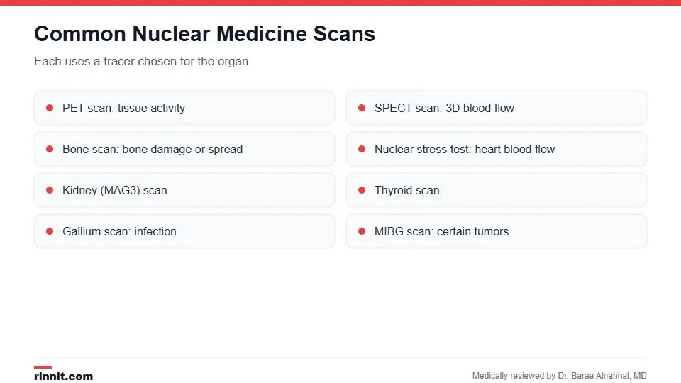
Each scan uses a tracer chosen for the organ being studied.
Scan | What it shows |
Tissue activity, often for cancer, heart and brain | |
3D images of blood flow and organ function | |
Bone damage, infection or cancer spread | |
Blood flow to the heart muscle | |
How the kidneys filter and drain |
Sources: [1]
How to prepare
Preparation depends on the scan. You may need to fast for several hours or adjust medicines.
Tell your provider about allergies and all medicines and supplements.
Tell your provider if you're pregnant, might be pregnant or are breastfeeding, because the test may not be safe.
Wear comfortable clothes and leave jewelry at home. Sources: [1]
What happens during the scan
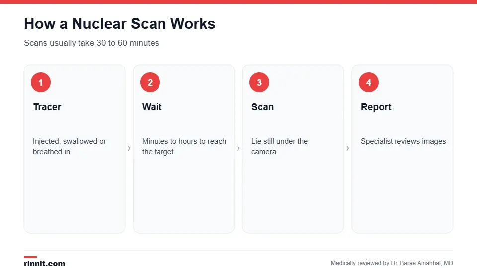
Scans usually take 30 to 60 minutes once the tracer is in place.
You receive the tracer, usually through an IV.
You wait while it travels to the area being studied. This can take minutes to hours; for a bone scan it takes several hours.
You lie still on a table while the camera or scanner takes pictures.
A nuclear medicine physician reviews the images and sends a report to your provider. Sources: [1]
Safety and side effects
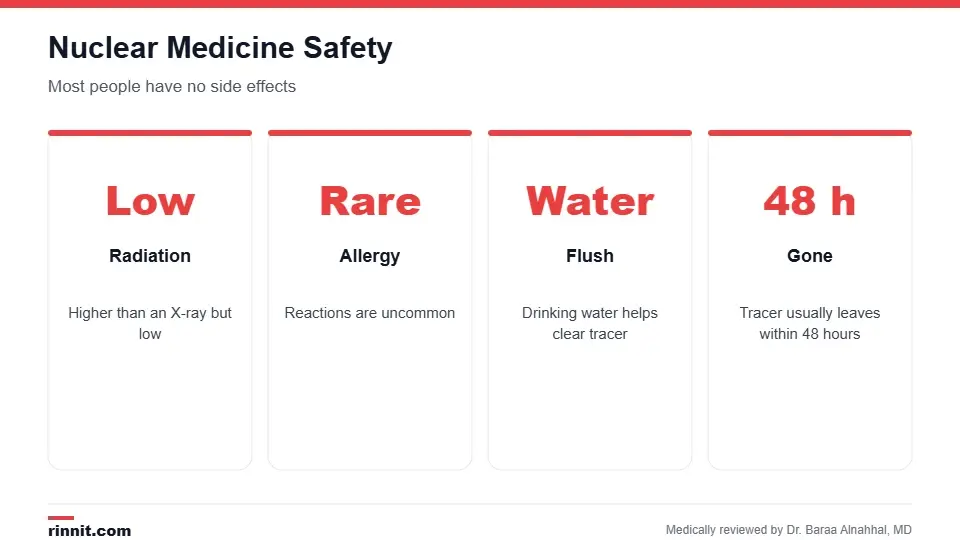
Most people have no side effects.
The radiation dose is low but higher than a standard X-ray.
Allergic reactions are rare.
The injection site may be sore or red.
You can usually eat, drink and return to normal activities right away. Sources: [1, 3]
Call your provider for swelling, itching or pain at the injection site, fever, or nausea and vomiting. Sources: [1]
Results
Results go to the provider who ordered the scan and are often ready within a few days. Depending on the findings, you may need more tests, such as a CT scan, MRI, blood tests or a biopsy. Sources: [1]
Conclusion
Nuclear medicine imaging shows how your body is working from the inside, often revealing problems other scans can't. It uses small, well-controlled amounts of radiation and is safe for most people. Ask your provider which scan you need and how to prepare.
Keep learning: read about the PET scan and the SPECT scan.
Frequently asked questions
What is nuclear medicine imaging?
Scans that use a small radioactive tracer to show how organs work.
What is a tracer?
A small amount of radioactive material that travels to a specific organ.
Is nuclear medicine safe?
Yes. It uses a low radiation dose and side effects are rare.
How long does a nuclear scan take?
Usually 30 to 60 minutes, plus waiting time for the tracer.
How long does the tracer stay in my body?
It's usually gone within 48 hours.
Can I have a nuclear scan while pregnant?
It may not be safe. Talk to your provider.
Can I breastfeed after a nuclear scan?
Ask your provider; you may need to pause for a time.
Will I feel anything?
Only the IV needle; the scan itself is painless.
Can I drive home?
Usually yes, unless you had a sedative.
When will I get results?
Usually within a few days.
Related Reading
Peptide Receptor Radionuclide Therapy (PRRT): How It Works explains PRRT for neuroendocrine tumors.
VQ Scan: Purpose, What to Expect and Results explains how a VQ scan checks for lung clots.
PSMA-Targeted Therapy (Pluvicto): How It Works and What to Expect explains PSMA-targeted therapy for advanced prostate cancer.
Gastric Emptying Study in Children (Solid-Meal GES): What to Expect explains the stomach emptying test for children.
Cisternogram Scan: Tracking Spinal Fluid Flow and Leaks explains the nuclear scan that tracks spinal fluid flow.
MUGA Scan: Measuring How Well Your Heart Pumps explains the nuclear scan that measures ejection fraction.
Cardiac PYP Scan: Testing for ATTR Cardiac Amyloidosis explains the nuclear scan used to diagnose ATTR cardiac amyloidosis.
Blood Volume Testing: How It Works and What Results Mean explains the nuclear test that measures total blood volume.
Liver-Spleen Scan: Nuclear Imaging of Liver and Spleen Function explains the nuclear scan that shows liver and spleen function.
Salivary Gland Scan (Parotid Scintigraphy): Uses and Results explains the nuclear scan that checks how well salivary glands work.
Parathyroid Scan (Sestamibi): Finding Overactive Glands Before Surgery explains the scan that finds overactive parathyroid glands before surgery.
PET Scan: Uses, Preparation, What to Expect and Results a common nuclear scan for cancer and heart disease.
SPECT Scan: How It Works, What It Shows and What to Expect a nuclear scan that creates 3D images.
Bone Scan: What It Shows, How It's Done, Results and Risks a nuclear scan for bone problems.
Nuclear Stress Test: How It Works, How to Prepare and What Results Mean a nuclear scan of heart blood flow.
Renal MAG3 Scan: Kidney Function and Drainage Test Explained a nuclear scan of kidney function.
Gallium Scan: What It Shows, How It Works and What to Expect a nuclear scan for infection and inflammation.
References
Cleveland Clinic. Nuclear Medicine & Imaging. Cleveland Clinic. Last updated December 18, 2025. https://my.clevelandclinic.org/health/diagnostics/4902-nuclear-medicine-imaging
Fazel R, Krumholz HM, Wang Y, et al. Exposure to low-dose ionizing radiation from medical imaging procedures. New England Journal of Medicine. 2009;361(9):849-857. PMID 19710483. https://doi.org/10.1056/NEJMoa0901249
Schreuder N, Jacobs NA, Jager PL, et al. Patient-reported adverse events of radiopharmaceuticals: a prospective study of 1002 patients. Drug Safety. 2021;44(2):211-222. PMID 33094442. https://doi.org/10.1007/s40264-020-01006-2
Medical disclaimer
This article is for general education only and does not replace advice from a qualified healthcare professional. Talk to your doctor about your own diagnosis and treatment, and seek emergency care right away for severe or sudden symptoms.



Comments