VQ Scan: Purpose, What to Expect and Results

Updated: 2 days ago
Written and medically reviewed by Dr. Baraa Alnahhal, MD · Last reviewed: November 2026 · How we review
Quick Answer
A VQ scan (ventilation-perfusion scan) is a two-part imaging test that measures airflow (ventilation) and blood flow (perfusion) in the lungs. It's most often used to check for a blood clot in the lungs (pulmonary embolism). It takes 30 to 60 minutes and uses no contrast dye and less radiation than a CT scan.
What the research says
VQ scans safely ruled out clots: in a trial of 1,417 people with suspected pulmonary embolism, when the test ruled out a clot, only 1.0% of the VQ scan group and 0.4% of the CT group had a clot over 3 months, though CT found more clots (19.2% vs. 14.2%) (Anderson DR et al., JAMA, 2007) [2]
VQ SPECT/CT was highly accurate: a systematic review found VQ SPECT/CT had a pooled sensitivity of 97.6% versus 82.0% for CT angiography, with accuracy of 96.5% versus 88.6% (Hess S et al., Seminars in Thrombosis and Hemostasis, 2016) [3]
The evidence on VQ SPECT is still limited: a systematic review found 13 diagnostic accuracy studies and one outcome study; 11 of the accuracy studies were at high risk of bias, so the exact performance of VQ SPECT for pulmonary embolism is still uncertain (Le Roux PY et al., Journal of Thrombosis and Haemostasis, 2020) [4]
VQ scans and CT are both options in pregnancy: a 2025 clinical review found that CT pulmonary angiography and VQ scans have comparable safety and effectiveness for suspected pulmonary embolism in pregnancy (Hammache M et al., Chest, 2025) [5]
Key takeaways
A VQ scan checks airflow and blood flow in your lungs.
It's mainly used to find a pulmonary embolism.
It uses a small amount of radioactive tracer but no contrast dye.
The test takes 30 to 60 minutes.
Results usually come back within 24 hours.
What is a VQ scan?
A VQ scan is also called a ventilation-perfusion scan, lung scan or lung scintigraphy. It's a nuclear medicine test. "V" stands for ventilation, and "Q" stands for perfusion. Sources: [1]
Why it's done

It's a good option for people who can't have contrast dye.
Diagnosing a pulmonary embolism, the most common reason.
Diagnosing some types of pulmonary hypertension.
Checking lung function after a lung transplant.
Evaluating your lungs before surgery to remove part of a lung.
Checking how well treatment for COPD is working. Sources: [1]
It's useful if you're allergic to contrast dye or have kidney problems. It uses less radiation than a CT angiogram. Sources: [1]
How to prepare
No special preparation is needed.
Tell your provider if you're pregnant, might be pregnant or are breastfeeding.
Let them know if you can't lie or sit still for several minutes.
You'll usually have a chest X-ray 1 to 24 hours before. Sources: [1]
What happens during the test

Each part takes about 15 minutes.
Ventilation: You breathe through a mask or mouthpiece connected to a machine with tracer while a camera takes pictures.
You may be repositioned for different views.
Perfusion: Right after, tracer is injected into a vein through an IV.
The same camera takes pictures of blood flow in your lungs. Sources: [1]
Each part takes about 15 minutes, and the whole test takes 30 to 60 minutes. Sources: [1]
Risks
A small amount of radiation that leaves your body within hours to days.
Both VQ scans and CT use low radiation in pregnancy. Guidelines often prefer a VQ scan when your chest X-ray is normal.
Allergic reactions are rare and usually mild.
Discomfort from the needle or staying still. Sources: [1]
After the test
The inhaled tracer leaves your lungs as you breathe, and the injected tracer leaves in your urine and stool within hours or days. If you're breastfeeding, ask your provider about feeding for a day or two afterward. Sources: [1]
Understanding results
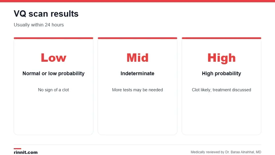
Results usually come back within 24 hours.
Result | What it means |
Normal or low probability | No sign of a pulmonary embolism |
Intermediate or indeterminate | More tests may be needed |
High probability | A pulmonary embolism is likely; your provider discusses treatment |
If air and blood flow images don't match, it may point to a clot. V/Q mismatch can have other causes too. Sources: [1, 2, 3]
VQ scan vs. CT scan
Feature | VQ scan | CT scan |
What the camera sees | Radioactive tracer | Contrast dye |
Contrast dye | Not used | Used |
Radiation | Less than a CT scan | More than a VQ scan |
Contrast allergy or kidney problems | Safe option | May not be suitable |
Both tests take pictures of your lungs to look for blood clots, but they work differently. Your provider picks the one that fits your situation. Sources: [1, 5]
Conclusion
A VQ scan is a low-risk, noninvasive way to check for blood clots in the lungs, especially if you can't have contrast dye. Results usually come quickly, so you and your provider can plan next steps.
Keep learning: read about pulmonary embolism and nuclear medicine imaging.
Frequently asked questions
What is a VQ scan?
A test that measures airflow and blood flow in your lungs.
What does a VQ scan check for?
Most often a blood clot in the lungs.
Is a VQ scan safe?
Yes. It uses a small amount of radiation.
Does a VQ scan use contrast dye?
No.
How long does a VQ scan take?
About 30 to 60 minutes.
Do I need to prepare?
No special preparation is needed.
Can I have a VQ scan if I'm pregnant?
Tell your provider. Guidelines often prefer a VQ scan in pregnancy when your chest X-ray is normal, while CT may be used in other cases.
When will I get results?
Usually within 24 hours.
Is a VQ scan better than a CT scan?
It uses less radiation and no contrast; CT may find more clots.
What does a high probability result mean?
A pulmonary embolism is likely.
Related Reading
Pulmonary Embolism Guide: Symptoms, Causes, Diagnosis, and Treatment the main condition a VQ scan finds.
Ventilation-Perfusion (V/Q) Mismatch: Causes, Symptoms, and Care what a mismatch means.
CT Angiogram (CTA): What It Shows, How to Prepare and What to Expect another test for lung clots.
Nuclear Medicine Imaging: How Tracer Scans Work, Safety and Results how tracer imaging works.
References
Cleveland Clinic. VQ Scan: Purpose, Procedure, Risks, Recovery & Results. Cleveland Clinic. Last updated July 7, 2026. https://my.clevelandclinic.org/health/treatments/17627-vq-scan
Anderson DR, Kahn SR, Rodger MA, et al. Computed tomographic pulmonary angiography vs ventilation-perfusion lung scanning in patients with suspected pulmonary embolism: a randomized controlled trial. JAMA. 2007;298(23):2743-2753. PMID 18165667. https://doi.org/10.1001/jama.298.23.2743
Hess S, Frary EC, Gerke O, et al. State-of-the-art imaging in pulmonary embolism: ventilation/perfusion single-photon emission computed tomography versus computed tomography angiography. Semin Thromb Hemost. 2016;42(8):833-845. PMID 27764879. https://doi.org/10.1055/s-0036-1593376
Le Roux PY, Robin P, Tromeur C, et al. Ventilation/perfusion SPECT for the diagnosis of pulmonary embolism: a systematic review. J Thromb Haemost. 2020;18(11):2910-2920. PMID 33433051. https://doi.org/10.1111/jth.15038
Hammache M, Simard C, Hamel S, et al. Diagnosing pulmonary embolism during pregnancy. Chest. 2025;168(4):1007-1017. PMID 40404047. https://doi.org/10.1016/j.chest.2025.05.014
Medical disclaimer
This article is for general education only and does not replace advice from a qualified healthcare professional. Talk to your doctor about your own diagnosis and treatment, and seek emergency care right away for severe or sudden symptoms.


Comments