CT Angiogram (CTA): What It Shows, How to Prepare and What to Expect

Updated: 2 days ago
Written and medically reviewed by Dr. Baraa Alnahhal, MD · Last reviewed: November 2026 · How we review
Quick Answer: What is a CT angiogram?
A CT angiogram (CTA) is a CT scan that uses contrast dye to make the blood vessels show up clearly, creating detailed 3D images. Providers use it to find blocked or narrowed arteries, aneurysms, tears in the aorta and blood clots in the lungs. You'll spend about 20 to 60 minutes in the testing room. [1]
What the research says
A heart CTA can help prevent heart attacks. In the SCOT-HEART trial of 4,146 people with stable chest pain, adding a coronary CT angiogram to standard care lowered the 5-year rate of death from coronary heart disease or heart attack from 3.9% to 2.3%, without increasing invasive procedures. (SCOT-HEART Investigators, The New England Journal of Medicine, 2018) [2]
CTA can safely replace a catheter test for many people. In a trial of 3,561 patients with stable chest pain, major cardiovascular events were similar after starting with CT (2.1%) or with invasive coronary angiography (3.0%), but major procedure-related complications were far less common with CT (0.5% vs 1.9%). (DISCHARGE Trial Group, The New England Journal of Medicine, 2022) [3]
It's highly sensitive for important heart artery narrowing. A meta-analysis of 77 studies found coronary CT angiography detected functionally significant blockages with 88% sensitivity, the highest of the noninvasive tests compared, which makes a normal CTA good at ruling out important coronary artery disease. (Pontone et al., International Journal of Cardiology, 2020) [4]
Key takeaways
A CT angiogram uses contrast dye and a CT scanner to make 3D images of your blood vessels. [1]
It can find narrowed or blocked arteries, aneurysms, aortic tears, lung clots and vessel injuries. [1]
The dye goes in through an IV, and you may feel a brief, harmless warm feeling. [1]
You're in the scanner for only 1 to 2 minutes and can resume normal activities right away. [1]
The main risks are an allergic reaction to the dye and a small dose of radiation. [1]
What is a CT angiogram?
A CT angiogram combines a computed tomography (CT) scan with contrast dye injected into a vein. The dye makes your blood vessels stand out brightly on the images, so your provider can see how blood flows and spot problems. [1]
The scanner takes hundreds of pictures that are combined into detailed 3D images of your blood vessels and the tissue around them. [1]
When a CT angiogram looks at the arteries of your heart, it's called a coronary CT angiogram (CCTA). [1]
What does a CT angiogram show?
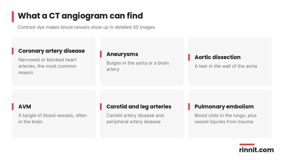
A CT angiogram can image arteries throughout your body, including the arteries of your heart, your aorta, the arteries in your brain and neck (carotid arteries), and those in your arms and legs. It can also look at your lungs for blood clots and at areas injured in chest, neck or head trauma. [1]
It helps diagnose: [1]
Narrowing or blockage of the heart arteries (coronary artery disease), the most common reason for the test
An aneurysm, a bulge in the wall of the aorta or a brain artery
Aortic dissection, a tear in the wall of the aorta
Arteriovenous malformation (AVM), a tangle of blood vessels, often in the brain
Carotid artery disease and peripheral artery disease
Pulmonary embolism, a blood clot in the lungs
Spontaneous coronary artery dissection (SCAD)
Blood vessel injuries from trauma
It's also used to plan procedures such as heart valve replacement, bypass surgery or stent placement. [1]
How do you prepare for a CT angiogram?
The provider who orders your test will give you specific instructions, including how long to fast and whether to pause any medicines. Tell them: [1]
Every medicine you take
Your medical history, including heart or kidney conditions and allergies
If you've ever had a reaction to contrast dye
If you're pregnant or breastfeeding
If you've reacted to contrast dye before, your provider may give you medicine beforehand or suggest a different test. On the day of the scan, you'll change into a hospital gown and remove jewelry and other metal objects. [1]
What happens during a CT angiogram?

You lie flat on the exam table. Pillows or straps can help you stay comfortable and still.
A provider places an IV in your hand or arm for the contrast dye.
For heart scans, EKG patches are placed on your chest so the images can be timed with your heartbeat.
The technologist moves to another room and talks with you through an intercom.
The table slides through the ring-shaped scanner as it takes hundreds of images. You may be asked to hold your breath or stay very still.
When the scan is done, the IV is removed and a bandage is placed.
You don't need anesthesia, and you stay awake. You may feel a brief, harmless warm feeling when the dye goes in and hear buzzing or clicking from the machine. The scanner is open and feels less closed-in than an MRI. [1]
CT angiogram at a glance
Item | Typical detail |
Time in the testing room | About 20 to 60 minutes, including prep |
Time in the scanner | About 1 to 2 minutes (you may go through more than once) |
Anesthesia | None; you stay awake |
What you may feel | A brief warm feeling from the dye |
Recovery | Return to normal activities right away |
Who reads the images | A radiologist |
Sources: [1]
CT angiogram vs. conventional angiogram
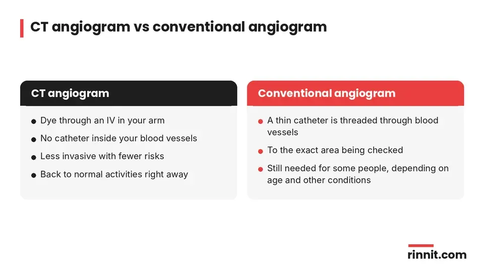
A conventional angiogram threads a thin tube (catheter) through your blood vessels to the area being checked. A CT angiogram doesn't need a catheter, so it's less invasive and has fewer risks. Depending on your age and other conditions, a CT angiogram may not be right for you, and you may still need a conventional angiogram. [1]
What are the risks of a CT angiogram?
The main risks are: [1]
An allergic reaction to the contrast dye. Most people have no problems, but reactions can happen, so tell your provider about any past reaction.
Radiation exposure. One CT angiogram gives a very small dose of radiation. Radiation that adds up over a lifetime may slightly raise your cancer risk, but the benefits usually outweigh the risks.
What happens after a CT angiogram?
You can go back to your normal activities right away. Drink plenty of water and other fluids to help flush the dye out of your body. [1]
A radiologist reviews the images and sends the findings to your provider, who will go over the results with you. Results can show plaque buildup, aneurysms and other problems, and you may need follow-up visits or more tests. Call your provider if you have questions about preparing for the test, your results or treatment. [1]
Questions to ask your provider
Why do I need a CT angiogram, and which vessels will it check?
Do I need to fast or pause any medicines?
What if I've had a reaction to contrast dye before?
How much radiation will I get?
When will I get my results, and who will explain them?
Conclusion
A CT angiogram gives your provider a detailed 3D view of your blood vessels using contrast dye and a CT scanner. It's one of the main ways to find blocked heart arteries, aneurysms, aortic tears and blood clots in the lungs, and it helps plan procedures such as stents and valve replacement.
Compared with a conventional angiogram, it's less invasive and has fewer risks. You'll be awake, the scan itself takes only a minute or two, and you can return to your day as soon as it's over.
Keep learning: Read our guides to the CT scan, the angiogram, the cardiac CT scan and cardiac catheterization. For more on this test, see RadiologyInfo.org: CT Angiography.
FAQ
What is a CT angiogram?
A CT angiogram is a CT scan with contrast dye that makes 3D images of your blood vessels and the tissue around them. [1]
What does a CT angiogram show?
It can show narrowed or blocked arteries, aneurysms, aortic dissection, AVMs, carotid and peripheral artery disease, lung clots and vessel injuries. [1]
How long does a CT angiogram take?
About 20 to 60 minutes in the testing room, with only 1 to 2 minutes inside the scanner. [1]
Is a CT angiogram painful?
No. You may feel a brief, harmless warm feeling when the dye goes in. [1]
Do I need to fast before a CT angiogram?
Your provider will tell you how long to fast and whether to pause any medicines. [1]
Am I awake during a CT angiogram?
Yes. No anesthesia is needed. [1]
What are the risks of a CT angiogram?
The main risks are an allergic reaction to the contrast dye and a small dose of radiation. [1]
How is a CT angiogram different from a regular angiogram?
A CT angiogram doesn't need a catheter threaded through your blood vessels, so it's less invasive and has fewer risks. [1]
What should I do after a CT angiogram?
Return to your normal activities and drink plenty of fluids to help flush out the dye. [1]
Who reads a CT angiogram?
A radiologist reviews the images and sends the findings to your provider, who explains the results. [1]
Related Reading
VQ Scan: Purpose, What to Expect and Results explains how a VQ scan checks for lung clots.
Learn more in our guide to Coronary Artery Disease: Symptoms, Causes, Diagnosis, Treatment and Prevention — What You Need to Know.
Learn more in our guide to Aortic Aneurysm: Types, Causes, Dissection Risk & Complete Guide.
Learn more in our guide to Aortic Dissection: Emergency Warning Signs, Types, Diagnosis & Treatment.
Learn more in our guide to Brain Aneurysm: Symptoms, Causes, and Treatment.
Learn more in our guide to Brain AVM (Arteriovenous Malformation): Symptoms, Causes, and Treatment.
Learn more in our guide to Carotid Artery Disease: The Silent Stroke Risk in Your Neck, Explained.
Learn more in our guide to Peripheral Artery Disease (PAD): Symptoms, Causes, Diagnosis & Treatment Options.
Learn more in our guide to Pulmonary Embolism Guide: Symptoms, Causes, Diagnosis, and Treatment.
Learn more in our guide to Spontaneous Coronary Artery Dissection (SCAD) Guide: Symptoms, Causes, Diagnosis, and Treatment.
References
Cleveland Clinic. CT Angiogram: Procedure & Purpose. Cleveland Clinic. Last updated May 15, 2025. https://my.clevelandclinic.org/health/diagnostics/ct-angiogram
SCOT-HEART Investigators; Newby DE, Adamson PD, Berry C, et al. Coronary CT Angiography and 5-Year Risk of Myocardial Infarction. N Engl J Med. 2018;379(10):924-933. PMID 30145934. https://doi.org/10.1056/NEJMoa1805971
DISCHARGE Trial Group; Maurovich-Horvat P, Bosserdt M, Kofoed KF, et al. CT or Invasive Coronary Angiography in Stable Chest Pain. N Engl J Med. 2022;386(17):1591-1602. PMID 35240010. https://doi.org/10.1056/NEJMoa2200963
Pontone G, Guaricci AI, Palmer SC, et al. Diagnostic performance of non-invasive imaging for stable coronary artery disease: A meta-analysis. Int J Cardiol. 2020;300:276-281. PMID 31748186. https://doi.org/10.1016/j.ijcard.2019.10.046
Medical disclaimer
This article is for general education only and does not replace advice from a qualified healthcare professional. Talk to your doctor about your own diagnosis and treatment, and seek emergency care right away for severe or sudden symptoms.



Comments