MUGA Scan: Measuring How Well Your Heart Pumps

Updated: 2 days ago
Written and medically reviewed by Dr. Baraa Alnahhal, MD · Last reviewed: November 2026 · How we review
Quick Answer
A MUGA scan (multigated acquisition scan) is a nuclear heart imaging test that shows how well the heart pumps by measuring the ejection fraction (EF), the percentage of blood pumped with each beat. It's used to check for heart failure and monitor the heart during cancer treatments that can harm it. A typical EF is 50% to 75%.
What the research says
It helps prevent chemo heart damage: in 1,487 people treated with doxorubicin, using MUGA results to guide treatment cut heart failure in high-risk patients from 20.8% to 2.9% (Schwartz RG et al., Am J Med, 1987) [2]
It's a reliable measure: in 50 people on trastuzumab, MUGA ejection fraction matched cardiac MRI closely, while standard 2D echocardiography matched poorly (Walker J et al., J Clin Oncol, 2010) [3]
Key takeaways
A MUGA scan measures how well your heart pumps.
It reports your ejection fraction; 50% to 75% is typical.
It's often used to monitor the heart during cancer treatment.
A tracer is injected and pictures are synced to your heartbeat.
The test takes 1 to 3 hours.
What is a MUGA scan?
"Multigated" means the camera's images are timed to your heartbeat using EKG electrodes. Pictures from hundreds of beats are combined to show your heart's lower chambers filling and emptying. Because it counts tracer directly, MUGA gives a very consistent EF measurement, which is useful for repeated tests over time. An echocardiogram or cardiac MRI can also measure EF. Sources: [1]
Why it's done

MUGA tracks how well the heart pumps over time.
Check for heart failure in people with chest pain, dizziness, fatigue, shortness of breath or swelling.
Check for heart damage before, during or after cancer treatment, including chemotherapy, immunotherapy, chest radiation or a bone marrow transplant. Sources: [1]
How to prepare
Avoid eating and drinking for up to 6 hours beforehand, as directed.
You may need to avoid caffeine for a period.
Tell your provider about all medicines; you may need to stop nitrates or digoxin.
Tell your provider if you're pregnant or breastfeeding. Sources: [1]
What happens during the test
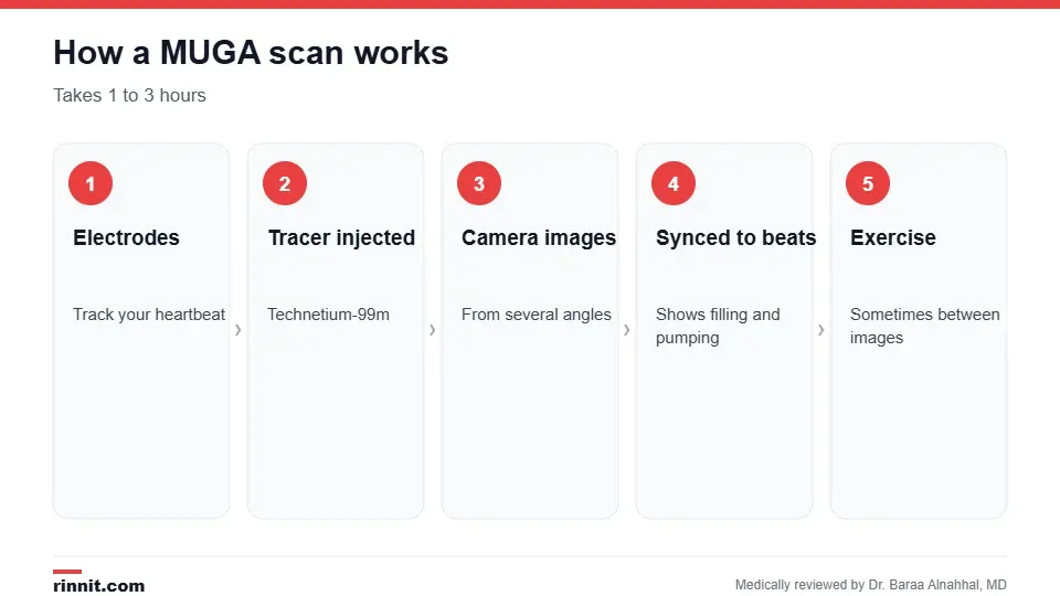
The test takes 1 to 3 hours.
Electrodes are attached to your chest to track your heartbeat.
A small amount of technetium-99m is injected into a vein.
You lie under a camera that takes pictures from several angles.
In some cases, you walk on a treadmill or exercise between images to see how your heart responds to stress. Sources: [1]
Risks
MUGA scans are safe for most people. The tracer usually leaves your body within 24 hours. People who are pregnant or breastfeeding typically shouldn't have the test. You can resume usual activities, including driving, right away. Drink plenty of fluids for a day or two. Sources: [1]
What the results mean

Your provider reviews results and next steps.
Ejection fraction | What it may mean |
50% to 75% | Typical pumping |
Below 50% | Weakened pumping, such as heart failure or damage from cancer treatment |
Above 75% | May point to a heart problem |
During cancer treatment, a drop in EF may lead your oncologist to adjust or pause the treatment. Research shows that using serial MUGA scans to guide doxorubicin chemotherapy sharply reduced heart failure. Sources: [1, 2, 3]
Conclusion
A MUGA scan gives a precise, repeatable measure of your heart's pumping strength. It's especially valuable for tracking heart health during cancer treatment and for diagnosing heart failure.
Keep learning: read about the echocardiogram and the nuclear stress test.
Frequently asked questions
What is a MUGA scan?
A nuclear test that measures how well your heart pumps.
What does MUGA stand for?
Multigated acquisition.
What is a normal ejection fraction?
About 50% to 75%.
Why is it used during chemotherapy?
Some cancer treatments can weaken the heart.
How long does it take?
1 to 3 hours.
Is it safe?
Yes, for most people; the tracer leaves within 24 hours.
Can I eat beforehand?
You may need to fast for up to 6 hours.
Does it hurt?
Only the IV needle.
Can I drive afterward?
Yes.
How is MUGA different from an echocardiogram?
MUGA uses a tracer; an echo uses ultrasound.
Related Reading
Echocardiogram: Types, What to Expect, Results and Risks ultrasound of the heart.
Heart MRI (Cardiac MRI): What It Shows, How to Prepare, What Happens and Results detailed heart imaging with MRI.
Nuclear Stress Test: How It Works, How to Prepare and What Results Mean a nuclear test of heart blood flow.
Heart Failure: Symptoms, Causes & Treatment Guide a condition MUGA helps assess.
Nuclear Medicine Imaging: How Tracer Scans Work, Safety and Results an overview of tracer-based tests.
References
Cleveland Clinic. Multigated Acquisition Scan (MUGA). Cleveland Clinic. Last updated June 1, 2022. https://my.clevelandclinic.org/health/diagnostics/17247-multigated-acquisition-scan-muga
Schwartz RG, McKenzie WB, Alexander J, et al. Congestive heart failure and left ventricular dysfunction complicating doxorubicin therapy: seven-year experience using serial radionuclide angiocardiography. Am J Med. 1987;82(6):1109-1118. PMID 3605130. https://doi.org/10.1016/0002-9343(87)90212-9
Walker J, Bhullar N, Fallah-Rad N, et al. Role of three-dimensional echocardiography in breast cancer: comparison with two-dimensional echocardiography, multiple-gated acquisition scans, and cardiac magnetic resonance imaging. J Clin Oncol. 2010;28(21):3429-3436. PMID 20530277. https://doi.org/10.1200/JCO.2009.26.7294
Medical disclaimer
This article is for general education only and does not replace advice from a qualified healthcare professional. Talk to your doctor about your own diagnosis and treatment, and seek emergency care right away for severe or sudden symptoms.



Comments