Echocardiogram: Types, What to Expect, Results and Risks

Updated: 6 days ago
Written and medically reviewed by Dr. Baraa Alnahhal, MD · Last reviewed: November 2026 · How we review
Quick Answer: What is an echocardiogram?
An echocardiogram, often shortened to "echo" or called a heart ultrasound, is an imaging test that uses high-frequency sound waves to create pictures of your heart. It shows the heart's structure, how well it pumps and how blood flows through its chambers and valves. Most echoes are noninvasive and use no radiation [1].
What the research says
Stress echo detects blocked heart arteries with balanced accuracy. A meta-analysis of 104 studies with 16,824 people with symptoms found stress echocardiography had a pooled sensitivity of 81% and specificity of 85% for obstructive coronary artery disease, far better than an exercise test alone (66% and 61%). (Sonaglioni et al., Journal of Clinical Medicine, 2025) [2]
A standard chest echo can miss infection growths on the valves. Across 16 studies with 2,807 people suspected of having infective endocarditis, a transthoracic echo found vegetations with 61% sensitivity and 94% specificity, using a transesophageal echo as the reference. (Bai et al., Journal of the American Society of Echocardiography, 2017) [3]
Transesophageal echo is highly sensitive for valve vegetations. In a meta-analysis of 8 studies checked against surgical findings, transesophageal echo detected vegetations with 94% sensitivity versus 64% for cardiac CT, and found leaflet perforations with 81% versus 41% sensitivity. (Oliveira et al., Radiology: Cardiothoracic Imaging, 2020) [4]
Key takeaways
An echocardiogram is an ultrasound of the heart that shows its structure, pumping function and blood flow through the valves [1].
The four main types are transthoracic (TTE), transesophageal (TEE), exercise stress echo and dobutamine stress echo [1].
A TTE usually takes 40 to 60 minutes, while a TEE can take up to 90 minutes [1].
A TTE has no known risks; a TEE may leave a mild sore throat for a few days [1].
You need someone to drive you home after a TEE and should not drive for 24 hours because of sedation [1].
An echo can help find valve disease, cardiomyopathy, blood clots in the heart, infective endocarditis and other conditions [1].
What is an echocardiogram used for?
Heart doctors (cardiologists) order an echocardiogram to look closely at how your heart is built and how it works. The images show the size of the chambers, the movement of the valves and the way blood travels through the heart [1].
Your provider may recommend an echo to [1]:
Find the cause of symptoms you are having
Confirm a suspected diagnosis
Check how well your heart is working
Keep track of a known condition, such as heart valve disease
Plan for heart surgery
See how well a heart surgery worked
What conditions can an echocardiogram detect?
An echo can reveal problems that need treatment now or monitoring over time. The conditions it can help identify include [1]:
Blood clots inside the heart chambers
Congenital heart disease (heart differences present from birth)
Cardiomyopathy (disease of the heart muscle)
Heart tumors
Infective endocarditis (infection of the heart's inner lining or valves)
Pericarditis (inflammation of the sac around the heart)
Thoracic aortic aneurysm (a bulge in the chest part of the aorta)
Heart valve disease

These findings guide whether you need treatment, further testing or regular follow-up echoes.
How does an echocardiogram work?
An echocardiogram works like other ultrasound scans. A handheld device called a transducer sends high-frequency sound waves into your body, and the echoes that bounce back from your heart are turned into moving images on a screen [1].
Because it relies on sound waves, an echo does not use any radiation [1]. It is also commonly used to monitor a known condition, such as valve disease [1].
What imaging techniques are used during an echo?
During one test, the team may use several imaging methods to answer different questions about your heart [1].
Technique | What it shows |
Two-dimensional (2D) imaging | The most common view; flat, moving pictures of the heart's structures |
Three-dimensional (3D) imaging | A three-dimensional view of the heart's structures |
Doppler ultrasound | The speed and direction of blood flow through the heart and valves |
Strain imaging | How the heart muscle moves as it squeezes and relaxes |
Contrast imaging | Dye injected into a vein to make heart structures easier to see |
Sources: [1]
What are the types of echocardiogram?
There are four main types of echocardiogram. Your provider chooses one based on what they need to see and whether you can exercise [1].
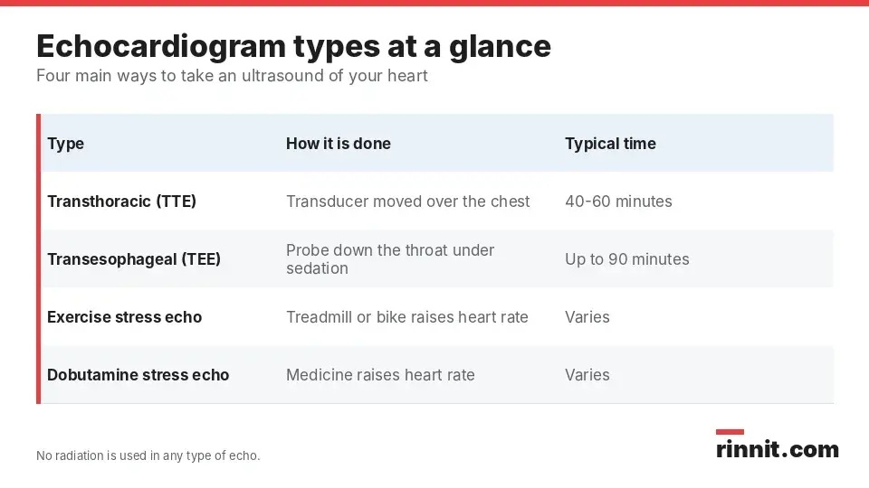
Each type gives a different view, so you may have more than one kind over time.
Transthoracic echocardiogram (TTE)
A TTE is the most common type. The transducer is moved over the surface of your chest, and the test is quick and painless [1].
Transesophageal echocardiogram (TEE)
For a TEE, a small ultrasound device is guided down your throat into your esophagus while you are sedated. From inside the body, a TEE produces higher-quality pictures and gives a clearer view of the back of the heart [1].
Research shows this closer view matters. When a standard chest echo looks for infection growths (vegetations) on heart valves, it misses a meaningful share that a TEE finds [3]. Compared with surgical findings, TEE detected vegetations with 94% sensitivity [4].
Exercise stress echocardiogram
An exercise stress echo checks how your heart works when it is under strain. You walk on a treadmill or pedal a stationary bike to raise your heart rate, and images are taken to show how the heart responds. It helps find the cause of symptoms that appear during physical activity [1].
Dobutamine stress echocardiogram
A dobutamine stress echo uses a medication, instead of exercise, to make your heart beat faster. It is an option if you are unable to exercise on a treadmill or bike [1].
Type | How it is done | Best for | Typical time |
Transthoracic (TTE) | Transducer on the chest | Quick, painless first look | 40-60 minutes |
Transesophageal (TEE) | Probe down the throat under sedation | Clearer images, back of the heart | Up to 90 minutes |
Exercise stress echo | Treadmill or bike raises heart rate | Symptoms during activity | Varies |
Dobutamine stress echo | Medication raises heart rate | People who cannot exercise | Varies |
Sources: [1]
How do you prepare for an echocardiogram?
Preparation depends on which type of echo you are having. Your provider will give you specific instructions for your test [1].
You may be asked to [1]:
Avoid caffeine, alcohol, tobacco or food for a set amount of time before the test
Keep taking your usual medicines unless your provider tells you to stop
Wear comfortable clothes and shoes if you are having an exercise stress echo
Arrange for someone to drive you home if you will receive sedation, as with a TEE
On the day of the test, you will usually undress from the waist up and change into a gown [1].
What happens during an echocardiogram?
A trained cardiac sonographer performs the echo. The steps are similar for most types, with a few extra steps for a TEE or stress echo [1].

Knowing the steps ahead of time can make the appointment feel more routine.
Get comfortable. You lie on an exam table. For a TTE, gel is placed on your chest to help the sound waves travel. For a TEE, you gargle a numbing liquid and receive sedation [1].
Heart rhythm monitoring. Small sticky patches called EKG electrodes are placed on your chest to track your heart rhythm during the test [1].
Imaging. The sonographer moves the transducer to capture pictures of your heart from different angles [1].
Follow instructions. You may be asked to stay still, hold your breath or breathe in a certain way at specific moments so the images come out clear [1].
After the test, you may be watched for about 30 minutes before you leave [1].
How long does an echocardiogram take?
A standard TTE usually takes 40 to 60 minutes. A TEE can take up to 90 minutes, and you may spend about 30 more minutes being monitored afterward [1].
Does an echocardiogram hurt?
A TTE is painless [1]. For a TEE, your throat is numbed and you are sedated, though you may have a mild sore throat for a few days afterward [1]. During a stress echo, the extra effort on your heart can bring on symptoms like chest pain or shortness of breath [1].
What are the risks of an echocardiogram?
An echocardiogram is a very safe test, and the level of risk depends on the type [1].
Type or step | Possible risks |
Transthoracic echo (TTE) | No known risks |
Transesophageal echo (TEE) | Mild sore throat for a few days; sedation means you cannot drive for 24 hours |
Stress echo | Chest pain or shortness of breath during the test |
Contrast dye | Possible allergic reaction |
Sources: [1]
Your care team will explain which of these apply to the type of echo you are having.
What do echocardiogram results mean?
After a TTE or stress echo, you can usually drive yourself home. After a TEE, you need a ride, and you should not drive for 24 hours because of the sedation [1].
The provider who ordered the test, often your heart doctor, will go over your results with you. It is best to review them together, in person or by phone, rather than trying to interpret the report on your own from your online health record [1].
What is a normal echocardiogram result?
A normal echo means your provider found no concerns about the structure or function of your heart [1]. An abnormal result may point to a condition such as valve disease, cardiomyopathy or a clot, which may need treatment right away or later [1].
How accurate is an echocardiogram?
Accuracy depends on the type of echo and the question being asked. For finding blocked heart arteries, stress echo showed a sensitivity of 81% and specificity of 85% across more than 16,000 people [2]. For valve infections, a TEE is more sensitive than a standard chest echo, so your doctor may order a TEE if a TTE does not give a clear answer [3, 4].
When should you call your doctor?
Contact the provider who ordered your echo if [1]:
You have questions about how to prepare
You notice a reaction or side effect after the test
You are worried about your results or unsure about next steps
Conclusion
An echocardiogram is one of the most useful tools for seeing how your heart is built and how well it works. It uses sound waves, not radiation, and the most common version is quick and painless [1].
Your provider will choose the type that best answers their question, from a simple chest echo to a TEE or stress test. Follow your preparation instructions, plan a ride if you will be sedated, and review the results with your doctor so you understand what comes next [1].
Keep learning: Read about Heart Valve Disease: Symptoms, Causes & Treatment, one of the most common reasons for an echo, and Endocarditis: Symptoms, Causes, Types, Diagnosis, Treatment and When to Seek Emergency Care — What You Need to Know.
FAQ
What is an echocardiogram?
An echocardiogram is an ultrasound of the heart. It uses sound waves to show the heart's structure, how it pumps and how blood flows through its valves [1].
Why would a doctor order an echocardiogram?
A doctor may order one to find the cause of symptoms, confirm a diagnosis, check heart function, monitor a condition like valve disease, or plan for and check the results of heart surgery [1].
What is the difference between a TTE and a TEE?
A TTE takes pictures from the surface of your chest. A TEE uses a small probe passed down your throat under sedation, which gives clearer images, especially of the back of the heart [1].
How do I prepare for an echocardiogram?
Follow your provider's instructions. You may need to avoid caffeine, alcohol, tobacco or food for a while, wear comfortable clothes for a stress test, and arrange a ride if you will be sedated [1].
Does an echocardiogram hurt?
A standard chest echo is painless. A TEE is done with numbing and sedation but may leave a mild sore throat for a few days [1].
How long does an echocardiogram take?
A TTE usually takes 40 to 60 minutes. A TEE can take up to 90 minutes, plus about 30 minutes of monitoring afterward [1].
Does an echocardiogram use radiation?
No. An echocardiogram uses sound waves to make images and does not involve radiation [1].
What does a normal echocardiogram mean?
A normal result means your provider found no concerns with your heart's structure or function [1].
What are the risks of an echocardiogram?
A TTE has no known risks. A TEE may cause a mild sore throat, a stress echo can trigger chest pain or shortness of breath, and contrast dye can cause an allergic reaction [1].
Can I drive after an echocardiogram?
You can usually drive after a TTE or stress echo. After a TEE, you need someone to drive you home, and you should not drive for 24 hours [1].
When should I call my doctor after an echocardiogram?
Call the provider who ordered your test if you have a reaction or side effect, or if you have questions about your results or next steps [1].
Related Reading
MUGA Scan: Measuring How Well Your Heart Pumps explains the nuclear scan that measures ejection fraction.
Heart Biopsy (Endomyocardial Biopsy): Procedure, Risks and Results explains the catheter procedure that takes tiny samples of heart muscle.
Echocardiogram With Strain: How Strain Imaging Spots Early Heart Damage explains the strain echo that spots early heart muscle changes.
Cardiac Imaging: Types of Heart Scans and What Each One Shows compares the main heart imaging tests and what each one shows.
BNP Test (B-Type Natriuretic Peptide): Normal Levels and What High BNP Means explains the blood test used to diagnose and track heart failure.
Dobutamine Stress Echocardiogram: How It Works, Preparation and Results explains the medicine-based stress test for people who can't exercise.
Heart Valve Disease: Symptoms, Causes & Treatment explains the valve problems an echo is most often used to find and monitor.
Endocarditis: Symptoms, Causes, Types, Diagnosis, Treatment and When to Seek Emergency Care — What You Need to Know covers the heart infection that a TTE or TEE can help detect.
Thoracic Aortic Aneurysm: Symptoms, Causes, Treatment & Complete Guide describes the chest aorta bulge that can show up on heart imaging.
What Is a Cardiac Tumor? A Complete Guide to Heart Tumors, Types, and Treatments explains the growths in or on the heart that an echo can reveal.
Adult Congenital Heart Disease: What It Is, How It's Diagnosed, and Why Lifelong Follow-Up Matters covers heart differences present from birth and why repeat echoes are part of lifelong care.
Exercise Stress Echocardiogram: Why It's Done, How to Prepare and What Results Mean explains the heart ultrasound done at rest and after exercise.
Bubble Study (Bubble Echo): Finding PFO, ASD and Lung Shunts explains the saline bubble ultrasound test that finds holes in the heart.
Hemodynamic Test: Checking Heart Output and Circulation for Fainting explains the heart output and circulation test used to find causes of fainting.
References
Cleveland Clinic. Echocardiogram: Types & What To Expect. Cleveland Clinic. Last updated October 6, 2025. https://my.clevelandclinic.org/health/diagnostics/16947-echocardiogram
Sonaglioni A, Polymeropoulos A, Baravelli M, et al. Diagnostic Accuracy of Exercise Stress Testing, Stress Echocardiography, Myocardial Scintigraphy, and Cardiac Magnetic Resonance for Obstructive Coronary Artery Disease: Systematic Reviews and Meta-Analyses of 104 Studies Published from 1990 to 2025. J Clin Med. 2025;14(17):6238. PMID 40943997. https://doi.org/10.3390/jcm14176238
Bai AD, Steinberg M, Showler A, et al. Diagnostic Accuracy of Transthoracic Echocardiography for Infective Endocarditis Findings Using Transesophageal Echocardiography as the Reference Standard: A Meta-Analysis. J Am Soc Echocardiogr. 2017;30(7):639-646.e8. PMID 28483353. https://doi.org/10.1016/j.echo.2017.03.007
Oliveira M, Guittet L, Hamon M, et al. Comparative Value of Cardiac CT and Transesophageal Echocardiography in Infective Endocarditis: A Systematic Review and Meta-Analysis. Radiol Cardiothorac Imaging. 2020;2(3):e190189. PMID 33778583. https://doi.org/10.1148/ryct.2020190189
Medical disclaimer
This article is for general education only and does not replace advice from a qualified healthcare professional. Talk to your doctor about your own diagnosis and treatment, and seek emergency care right away for severe or sudden symptoms.



Comments