Exercise Stress Echocardiogram: Why It's Done, How to Prepare and What Results Mean

Updated: 2 days ago
Written and medically reviewed by Dr. Baraa Alnahhal, MD · Last reviewed: November 2026 · How we review
Quick Answer
An exercise stress echocardiogram (stress echo) is an ultrasound of the heart taken at rest and again after you exercise on a treadmill or bike. It shows whether every part of the heart muscle gets enough blood when it works harder. It's used to check for coronary artery disease. Avoid caffeine for 24 hours. It uses no radiation.
What the research says
Stress echo is the most balanced non-invasive test: a meta-analysis of 104 studies and 16,824 people with suspected or known coronary artery disease found stress echo had 81% sensitivity and 85% specificity, compared with 66% and 61% for an exercise EKG alone, 82% and 74% for nuclear SPECT, and 83% and 89% for stress cardiac MRI (Sonaglioni A et al., Journal of Clinical Medicine, 2025) [2]
It works well in people with high blood pressure: across 25 studies and 3,812 hypertensive patients, stress echo detected significant coronary disease with 85% sensitivity and 81% specificity, outperforming nuclear perfusion imaging (81% and 64%) and exercise EKG (53% and 51%), and its accuracy was not reduced by a thickened heart muscle (Bytyci I et al., Archives of Medical Science, 2025) [3]
Key takeaways
Echo images at rest and right after exercise are compared to find heart muscle that doesn't move normally under stress.
Main use: diagnosing coronary artery disease; also valve disease, cardiomyopathy, heart failure and pulmonary hypertension.
Prepare: no caffeine for 24 hours, no food for a few hours, no tobacco that day, comfortable shoes.
About 10–15 minutes of exercise; about 1 hour in total; no radiation.
If you can't exercise, dobutamine or pacemaker pacing can raise the heart rate instead.
What is an exercise stress echocardiogram?
An echocardiogram uses sound waves to make moving pictures of the heart. A narrowed coronary artery may supply enough blood at rest but not during exercise, when the heart needs more oxygen. During a stress echo, the part of the heart wall fed by that artery squeezes less well under stress, and the change shows on the images. Sources: [1]

A stress echo compares heart images at rest and right after exercise.
Why is it done?
Chest pain or pressure, shortness of breath, dizziness or an irregular heartbeat that comes on with activity.
Checking known coronary disease, or results after a stent or bypass.
Assessing heart valve disease, cardiomyopathy, heart failure or pulmonary hypertension.
Checking fitness before surgery, for athletes, or before extreme activities such as diving or high altitude. Sources: [1]
The test is not done in people with an aortic dissection, active infection or inflammation of the heart (endocarditis, myocarditis, pericarditis), ongoing chest pain, a very recent heart attack, severe aortic stenosis or an uncontrolled rhythm problem. Sources: [1]
How do I prepare?
Avoid caffeine (coffee, tea, cola, chocolate, energy drinks) for 24 hours.
Don't eat or drink for a few hours before the test, as instructed.
Don't smoke or use tobacco that day.
Take your medicines as your provider directs; don't stop any, including heart medicines, unless told to.
Wear comfortable clothes and walking shoes. Sources: [1]
What happens during the test?
Electrode patches are placed on your chest and a blood pressure cuff on your arm.
You lie on your left side while a sonographer takes resting echo images.
You walk on a treadmill or pedal a bike, starting slowly and getting harder, for about 10–15 minutes, until you reach your target heart rate (based on age and fitness) or your symptoms appear.
Tell the team right away about chest, arm or jaw discomfort, shortness of breath or dizziness.
You lie back down immediately for the stress images (on a bike, images may be taken while you pedal).
You cool down and go home when your heart rate and blood pressure return to normal. Sources: [1]
The whole appointment takes about 1 hour. Sources: [1]
Stress echo vs other stress tests
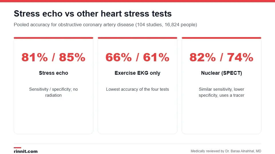
Pooled accuracy from 104 studies for obstructive coronary artery disease.
Test | How stress is shown | Sensitivity / specificity | Radiation |
Exercise EKG | Electrical changes on the EKG | 66% / 61% | None |
Exercise stress echo | Wall motion on ultrasound | 81% / 85% | None |
Nuclear stress test (SPECT) | Blood flow with an injected tracer | 82% / 74% | Yes |
Stress cardiac MRI | Blood flow and wall motion on MRI | 83% / 89% | None |
Stress echo offers the most balanced accuracy among widely available tests, without radiation. See also our guides to the exercise stress test and nuclear stress test. Sources: [2]
Understanding your results
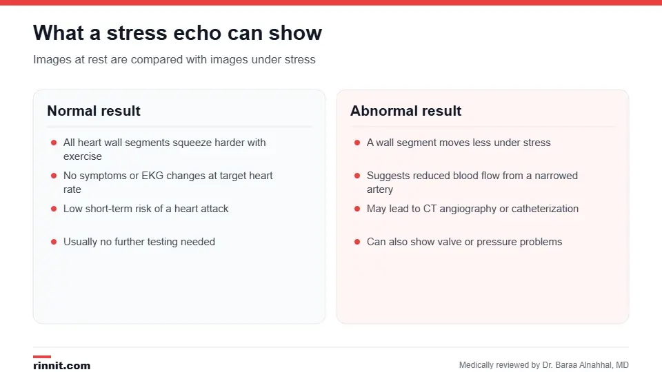
What a normal and an abnormal stress echo mean.
Your cardiologist compares how each segment of the heart wall moves at rest and under stress. A normal result means all segments squeeze harder with exercise. An abnormal result, where a segment moves less under stress, suggests reduced blood flow from a narrowed artery and may lead to coronary CT angiography or a coronary angiogram. The test can also reveal valve problems or high lung pressures. Sources: [1]
Risks
The test is safe and closely monitored. Most risks come from the heart condition being tested: exercise can trigger chest pain or an irregular rhythm, and the team stops the test if needed. Sources: [1]
Conclusion
An exercise stress echo shows how your heart performs when it has to work hard, with good accuracy and no radiation. Prepare by skipping caffeine for a day and wearing walking shoes, and report any symptoms during the test.
Keep learning: read about angina and the transthoracic echocardiogram.
Frequently asked questions
How long does a stress echo take?
About 1 hour in total, with 10–15 minutes of exercise.
Why can't I have caffeine before the test?
Caffeine affects heart rate and blood pressure and can interfere with the test. Avoid it for 24 hours.
What if I can't exercise?
A dobutamine stress echo uses medicine to raise your heart rate, or a pacemaker can be programmed to do it.
Is a stress echo better than a regular stress test?
It is more accurate than an exercise EKG alone: about 81% sensitivity and 85% specificity versus 66% and 61%.
Does a stress echo use radiation?
No. It uses ultrasound, unlike a nuclear stress test.
What does an abnormal result mean?
Part of the heart wall moved less under stress, suggesting a narrowed coronary artery. Further tests such as angiography may follow.
Should I take my heart medicines?
Follow your provider's instructions. Some medicines that slow the heart may be paused; don't stop any on your own.
What should I wear?
Comfortable clothing and walking shoes; you will change into a gown for the top half.
Is the test safe?
Yes. It is closely monitored and stopped if you develop concerning symptoms or rhythm changes.
Who performs the test?
A cardiac sonographer takes the images under the supervision of a physician, and a cardiologist interprets them.
Related Reading
Dobutamine Stress Echocardiogram: How It Works, Preparation and Results explains the medicine-based stress test for people who can't exercise.
Echocardiogram: Types, What to Expect, Results and Risks how heart ultrasound works and its main types.
Exercise Stress Test: Why It's Done, How to Prepare, What Happens and Results the EKG-only version of the exercise test.
Nuclear Stress Test: How It Works, How to Prepare and What Results Mean the tracer-based alternative to stress echo.
Angina: Symptoms, Causes, Treatment & Complete Guide the chest pain stress echo is often used to investigate.
Coronary Angiogram: Why It's Done, What to Expect and Recovery the next step when a stress echo is abnormal.
References
Cleveland Clinic. Exercise Stress Echocardiogram: Purpose and Procedure Details. Cleveland Clinic. Last updated July 8, 2022. https://my.clevelandclinic.org/health/diagnostics/16983-exercise-stress-echocardiogram
Sonaglioni A, Polymeropoulos A, Baravelli M, et al. Diagnostic accuracy of exercise stress testing, stress echocardiography, myocardial scintigraphy, and cardiac magnetic resonance for obstructive coronary artery disease: systematic reviews and meta-analyses of 104 studies published from 1990 to 2025. Journal of Clinical Medicine. 2025;14(17):6238. PMID 40943997. https://doi.org/10.3390/jcm14176238
Bytyci I, Bytyqi S, Abazi G, et al. Diagnostic accuracy of non-invasive investigations for coronary artery disease in hypertensive patients: a meta-analysis. Archives of Medical Science. 2025;21(1):167-178. PMID 40190327. https://doi.org/10.5114/aoms/188782
Medical disclaimer
This article is for general education only and does not replace advice from a qualified healthcare professional. Talk to your doctor about your own diagnosis and treatment, and seek emergency care right away for severe or sudden symptoms.



Comments