Transthoracic Echocardiogram (TTE): What It Shows, What to Expect and Results

Updated: 2 days ago
Written and medically reviewed by Dr. Baraa Alnahhal, MD · Last reviewed: November 2026 · How we review
Quick Answer: What is a transthoracic echocardiogram?
A transthoracic echocardiogram (TTE) is the most common type of echocardiogram. It uses sound waves from a wand moved across the chest to create moving pictures of the heart's chambers, valves and nearby blood vessels. It shows how well the heart pumps and helps diagnose heart failure and valve disease. It takes about an hour and uses no radiation. [1]
What the research says
Echocardiography often finds heart valve disease people did not know they had. When 2,500 adults aged 65 and older from primary care were screened with transthoracic echocardiography, 6.4% had previously undiagnosed moderate or severe valve disease, and about half had at least mild new valve changes. (d'Arcy et al., European Heart Journal, 2016) [2]
Contrast used during echocardiograms appears safe. A meta-analysis of 8 studies found no rise in deaths or heart attacks with ultrasound contrast agents; death occurred in 0.34% of people who received contrast versus 0.9% who did not. (Khawaja et al., The American Journal of Cardiology, 2010) [3]
Key takeaways
A TTE uses ultrasound to make moving images of your heart through your chest wall. [1]
It shows your heart's chambers, valves, pumping strength and blood flow. [1]
You can eat, drink and take your medicines as usual beforehand. [1]
The test takes about an hour and needs no sedation. [1]
There is no radiation, and you can drive yourself home afterward. [1]
What is a transthoracic echocardiogram?
A transthoracic echocardiogram is an ultrasound of your heart taken from outside your chest. High-frequency sound waves bounce off your heart tissue, and a computer turns the echoes into moving pictures on a screen that you and your provider can watch. A Doppler ultrasound may also be used to measure blood flow through your heart's chambers and valves. [1]
A primary care provider or cardiologist usually orders the test, and a sonographer, a technician trained in ultrasound, performs it and sends the results to your doctor. [1]
What does a TTE show?
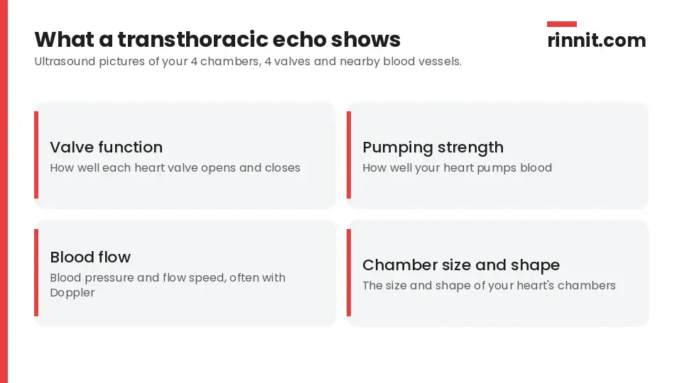
A TTE shows your heart's structure and how it works.
A TTE checks: [1]
How well your heart valves work
How well your heart pumps blood
Blood pressure and how fast blood flows through your heart
The size and shape of your heart's chambers
Why would you need a TTE?
Your provider may order a TTE to check your heart before or after a diagnosis or treatment, to find the cause of symptoms or to screen for heart conditions. [1]
Symptoms it can help explain
Chest pain
Swelling (edema)
A heart murmur
Shortness of breath
Sources: [1]
Conditions it can diagnose or monitor
Heart failure
Heart valve disease
Hypertrophic cardiomyopathy
Congenital heart conditions
Aortic aneurysm and aortic dissection
Blood clots
Heart tumors
Changes seen on an ECG
Sources: [1]
TTE vs. TEE: What is the difference?

A TEE may follow a TTE when clearer pictures are needed.
Feature | Transthoracic echo (TTE) | Transesophageal echo (TEE) |
How it's done | Wand moved across the outside of your chest | Thin scope passed down your esophagus |
Invasiveness | Noninvasive (or minimally invasive if contrast is injected) | Invasive |
When it's used | Most common first test | When clearer, more detailed pictures are needed after a TTE |
Sources: [1]
How do you prepare for a TTE?
No special preparation is needed. You can eat, drink and take your medicines as usual. [1]
What happens during a TTE?
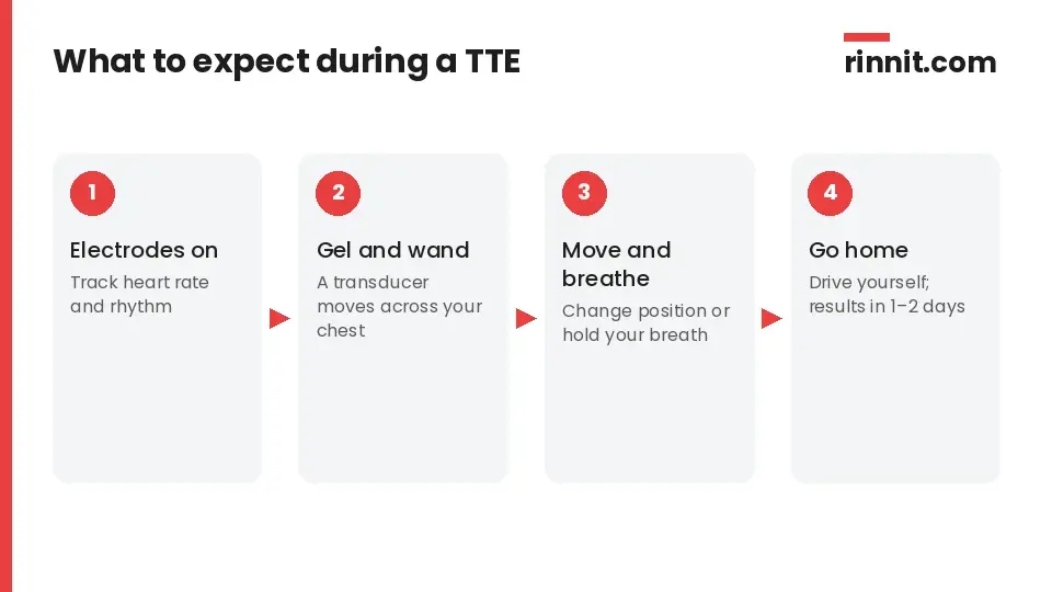
A TTE takes about an hour and uses no radiation.
You lie on an exam table, and electrodes are placed on your chest to track your heart rate and rhythm. A sonographer spreads gel on your skin to help the sound waves travel and moves a transducer (ultrasound wand) across your chest. You may be asked to hold still, change position or breathe in certain ways to get clearer pictures. [1]
Sometimes a provider injects a contrast agent into a vein to make blood flow through your heart easier to see. No sedation is needed. [1]
How long does a TTE take?
About an hour. [1]
Does a TTE hurt?
It should not hurt. If you receive contrast through an IV, you may feel a brief pinch. [1]
What happens afterward?
The technician removes the gel and electrodes, and you can go home right away, return to your normal activities and drive yourself. [1]
Are there risks?
A TTE has essentially no risks, because it uses no radiation or electrical current. [1]
When will you get your TTE results?
The sonographer sends a report to the provider who ordered the test, usually within a day or two. Your provider then explains the results, any diagnosis and your treatment plan, or whether you need more tests, such as a TEE. [1]
Conclusion
A transthoracic echocardiogram is a safe, painless and widely used way to see your heart at work. It shows your chambers, valves and blood flow without radiation, and it often guides the next step in diagnosing symptoms like chest pain, swelling or shortness of breath.
Ask your provider why they recommend the test and what your results mean for your heart health.
Keep learning: Read our guide to echocardiograms and their types or our guide to heart failure. For more background, see the MedlinePlus page on echocardiograms.
FAQ
What is a transthoracic echocardiogram (TTE)?
An ultrasound of your heart taken through your chest that shows its chambers, valves and blood flow. [1]
What does a TTE show?
How your valves work, how well your heart pumps, blood flow speed and the size and shape of your heart's chambers. [1]
Why would I need a TTE?
To find the cause of symptoms like chest pain, swelling, a murmur or shortness of breath, or to diagnose and monitor heart conditions. [1]
How do I prepare for a TTE?
No preparation is needed; eat, drink and take your medicines as usual. [1]
How long does a TTE take?
About an hour. [1]
Does a TTE hurt?
No, apart from a brief pinch if contrast is given through an IV. [1]
Is a TTE safe?
Yes; it uses no radiation or electrical current. [1]
What is the difference between TTE and TEE?
A TTE is done from outside your chest, while a TEE passes a scope down your esophagus for more detailed pictures. [1]
Can I drive after a TTE?
Yes; you can go home right away and drive yourself. [1]
When will I get my TTE results?
The report usually reaches your provider within a day or two. [1]
Related Reading
Heart Failure: Symptoms, Causes & Treatment Guide explains a condition a TTE helps diagnose and monitor.
Aortic Stenosis: Narrowed Heart Valve Symptoms, Causes & Treatment covers a valve disease commonly found on echocardiography.
What Is Mitral Valve Regurgitation? A Plain-Language Guide to This Common Heart Valve Problem describes a leaky valve a TTE can detect.
Heart Murmurs: What They Mean, When They're Harmless, and How They're Treated explains a finding that often leads to a TTE.
Congenital Heart Disease (CHD): Types, Symptoms, Treatment & Survival — Complete Guide covers heart defects a TTE helps diagnose.
Aortic Aneurysm: Types, Causes, Dissection Risk & Complete Guide describes an aorta problem a TTE can help find.
References
Cleveland Clinic. Transthoracic Echocardiogram (TTE). Cleveland Clinic. Last updated August 5, 2022. https://my.clevelandclinic.org/health/diagnostics/13477-echocardiogram-transthoracic-tte
d'Arcy JL, Coffey S, Loudon MA, et al. Large-scale community echocardiographic screening reveals a major burden of undiagnosed valvular heart disease in older people: the OxVALVE Population Cohort Study. Eur Heart J. 2016;37(47):3515-3522. PMID 27354049. https://doi.org/10.1093/eurheartj/ehw229
Khawaja OA, Shaikh KA, Al-Mallah MH. Meta-analysis of adverse cardiovascular events associated with echocardiographic contrast agents. Am J Cardiol. 2010;106(5):742-747. PMID 20723656. https://doi.org/10.1016/j.amjcard.2010.04.034
Medical disclaimer
This article is for general education only and does not replace advice from a qualified healthcare professional. Talk to your doctor about your own diagnosis and treatment, and seek emergency care right away for severe or sudden symptoms.



Comments