Cardiac Imaging: Types of Heart Scans and What Each One Shows

Updated: 3 days ago
Written and medically reviewed by Dr. Baraa Alnahhal, MD · Last reviewed: November 2026 · How we review
Quick Answer
Cardiac imaging is a group of tests that make pictures of the heart and nearby blood vessels. The main types are the echocardiogram, cardiac CT, cardiac MRI, nuclear stress tests, the coronary angiogram and the MUGA scan. They help screen for, diagnose and monitor problems such as coronary artery disease, heart failure and valve disease.
What the research says
Coronary CT can lower heart attack risk: in the SCOT-HEART trial of patients with stable chest pain, adding coronary CT angiography to standard care led to a significantly lower rate of death from coronary heart disease or nonfatal heart attack over 5 years, without a significant rise in invasive angiograms or stent procedures (SCOT-HEART Investigators (Newby DE et al.), New England Journal of Medicine, 2018) [2]
Anatomy-first and function-first testing gave similar outcomes: in the PROMISE trial of 10,003 people with symptoms of coronary artery disease, starting with CT angiography or with functional testing (exercise ECG, nuclear stress or stress echo) gave similar rates of death, heart attack and other major events over about 2 years (Douglas PS et al., New England Journal of Medicine, 2015) [3]
Key takeaways
Cardiac imaging includes echo, CT, MRI, nuclear tests, angiograms and MUGA scans.
Each test answers a different question about structure, blood flow or function.
Echo and MRI use no radiation; CT, angiograms and nuclear tests do.
Tell your provider about implants, metal, medicines and pregnancy.
Your provider chooses the test based on your symptoms and history.
What is cardiac imaging?
Cardiac imaging lets providers see your heart's chambers, valves, walls, muscle and blood vessels, and how blood flows through them. Some tests show structure, some show blood flow and some show how well the heart muscle pumps. Tests are sometimes combined, such as PET with CT. Sources: [1]
Types of cardiac imaging

Each test shows a different part of heart health.
Test | How it works | What it shows |
Echocardiogram | Ultrasound waves | Pumping, valves, walls, clots, holes in the heart |
Cardiac CT | X-rays combined into 3D images | Artery blockages, structure, procedure planning |
Cardiac MRI | Magnets and radio waves | Detailed structure, function, scarring and inflammation |
Nuclear stress test (PET, SPECT) | Radioactive tracer at rest and with stress | Blood flow to heart muscle, scar tissue |
Coronary angiogram | Catheter, dye and X-ray video | Blocked or narrowed coronary arteries |
MUGA scan | Radioactive tracer | Ejection fraction, often during chemotherapy |
Related tests include the cardiac PET scan, the calcium score test and the dobutamine stress echocardiogram. Sources: [1]
Why it's done
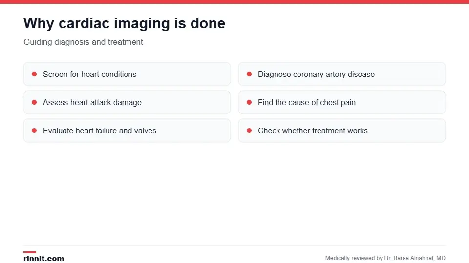
Imaging guides both diagnosis and treatment.
Screen for heart conditions.
Diagnose coronary artery disease, heart failure, valve disease, pericardial disease and congenital heart conditions.
See whether a heart attack happened and how much damage it caused.
Find the cause of chest pain or shortness of breath.
Check whether treatment is working. Sources: [1, 2, 3]
How to prepare
Avoid caffeine before stress tests and coronary angiograms.
Leave jewelry at home for CT and MRI, and wear metal-free clothing for MRI.
Tell your provider about all medical conditions, medicines and supplements.
Mention implanted devices such as pacemakers or mechanical valves, any metal in your body, and pregnancy. Sources: [1]
Radiation and risks
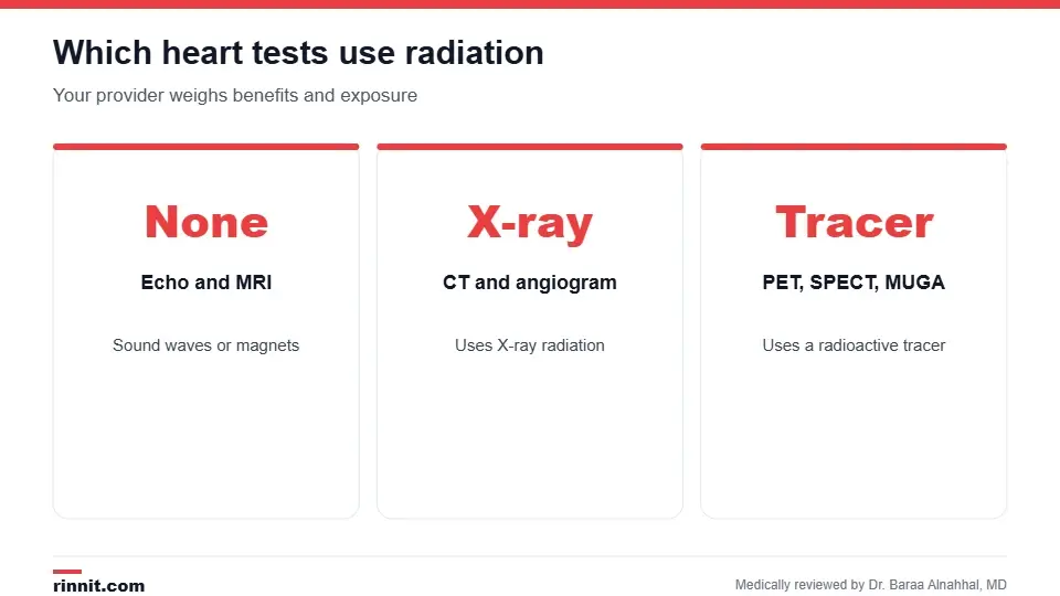
Your provider weighs the benefits and the radiation involved.
Radiation type | Tests |
None | Echocardiogram, cardiac MRI |
X-ray radiation | Cardiac CT, coronary angiogram |
Radioactive tracer | Nuclear stress tests (PET, SPECT), MUGA scan |
Cardiac imaging is generally safe because the tests are noninvasive or minimally invasive. Rarely, people react to the contrast dye used in some tests. Sources: [1]
Conclusion
Cardiac imaging gives your care team a clear view of your heart's structure, blood flow and pumping. Your provider will choose the test that best answers your question with the least risk.
Keep learning: read about the echocardiogram and cardiac MRI.
Frequently asked questions
What is cardiac imaging?
Tests that make pictures of your heart and blood vessels.
What is the most common cardiac imaging test?
The echocardiogram.
Which tests don't use radiation?
Echocardiograms and cardiac MRI.
Which tests use radiation?
Cardiac CT, coronary angiograms, nuclear stress tests and MUGA scans.
What does a coronary angiogram show?
Blocked or narrowed coronary arteries.
What is a MUGA scan used for?
Measuring ejection fraction, often during chemotherapy.
Do I need to avoid caffeine?
Yes, before stress tests and coronary angiograms.
Can I have an MRI with a pacemaker?
Tell your provider; some devices need special steps.
Is cardiac imaging safe?
Yes, most tests are noninvasive or minimally invasive.
How does my provider choose a test?
Based on your symptoms, history and what question needs answering.
Related Reading
Echocardiogram With Strain: How Strain Imaging Spots Early Heart Damage explains the strain echo that spots early heart muscle changes.
Echocardiogram: Types, What to Expect, Results and Risks the most common heart imaging test.
Cardiac CT Scan (Heart CT): Purpose, Preparation, Calcium Score and Risks detailed X-ray images of the heart and arteries.
Heart MRI (Cardiac MRI): What It Shows, How to Prepare, What Happens and Results radiation-free, detailed heart imaging.
Nuclear Stress Test: How It Works, How to Prepare and What Results Mean imaging blood flow to the heart muscle.
Coronary Angiogram: Why It's Done, What to Expect and Recovery the gold-standard look at the coronary arteries.
References
Cleveland Clinic. Cardiac Imaging: Types, Uses and Procedure Details. Cleveland Clinic. Last updated May 3, 2022. https://my.clevelandclinic.org/health/diagnostics/16836-cardiac-imaging
SCOT-HEART Investigators, Newby DE, Adamson PD, et al. Coronary CT angiography and 5-year risk of myocardial infarction. New England Journal of Medicine. 2018;379(10):924-933. PMID 30145934. https://doi.org/10.1056/NEJMoa1805971
Douglas PS, Hoffmann U, Patel MR, et al. Outcomes of anatomical versus functional testing for coronary artery disease. New England Journal of Medicine. 2015;372(14):1291-1300. PMID 25773919. https://doi.org/10.1056/NEJMoa1415516
Medical disclaimer
This article is for general education only and does not replace advice from a qualified healthcare professional. Talk to your doctor about your own diagnosis and treatment, and seek emergency care right away for severe or sudden symptoms.



Comments