Cardiac PET Scan: Heart Blood Flow Imaging, Prep and Results

Updated: 3 days ago
Written and medically reviewed by Dr. Baraa Alnahhal, MD · Last reviewed: November 2026 · How we review
Quick Answer
A cardiac PET scan is a nuclear imaging test that uses a small amount of radioactive tracer and a PET scanner to make 3D images showing how well blood flows to the heart muscle and how healthy the muscle cells are. It helps diagnose coronary artery disease. Fast for 4 hours and avoid caffeine for 24 hours beforehand.
What the research says
PET is highly sensitive for coronary artery disease: a meta-analysis of studies including 11,862 patients found PET stress imaging had a sensitivity of about 93% for significant coronary narrowing, higher than the 88% seen with SPECT, with similar specificity (Parker MW et al., Circ Cardiovasc Imaging, 2012) [2]
PET was the most accurate test head-to-head: in 208 people with suspected coronary disease who had CT angiography, SPECT and PET before invasive pressure testing, PET had the highest overall accuracy (85%) compared with CT angiography (74%) and SPECT (77%) (Danad I et al., JAMA Cardiol, 2017) [3]
Key takeaways
Shows blood flow and heart muscle health with a radioactive tracer.
Diagnoses coronary artery disease and checks heart damage after a heart attack.
Includes rest and medicine-induced stress images.
Fast 4 hours and avoid all caffeine for 24 hours.
Results usually in 24–48 hours.
What is a cardiac PET scan?
PET stands for positron emission tomography. A tracer injected into a vein travels to the heart, and the PET scanner detects it to create 3D images. Unlike tests that only show structure, PET shows what the heart cells are doing. Many centers use a combined PET/CT scanner. It's a type of nuclear stress test, similar to a SPECT scan, and related to a general PET scan. Sources: [1]
Why is it done?

Cardiac PET shows blood flow and heart muscle health.
Coronary artery disease and areas with poor blood flow.
Heart muscle damage after a heart attack.
Diseased heart muscle (cardiomyopathy).
Heart inflammation or infection, and cardiac sarcoidosis.
Whether a procedure such as a stent or bypass surgery would help, and whether current treatment is working. Sources: [1]
How do I prepare?

Caffeine can interfere with stress medicines.
Fasting: nothing but water for 4 hours before.
Caffeine: none for 24 hours, including coffee, tea, soda, chocolate, decaf products and some over-the-counter medicines.
Exercise and smoking: no strenuous exercise for 24 hours and no smoking on the day of the scan.
Medicines: theophylline and dipyridamole are usually stopped 48 hours before; ask about insulin and diabetes pills, and don't stop heart medicines without your provider's advice.
Special diet: for sarcoidosis or infection checks, you may follow a high-fat, low-carb diet for 24–48 hours. Sources: [1]
What happens during the scan?
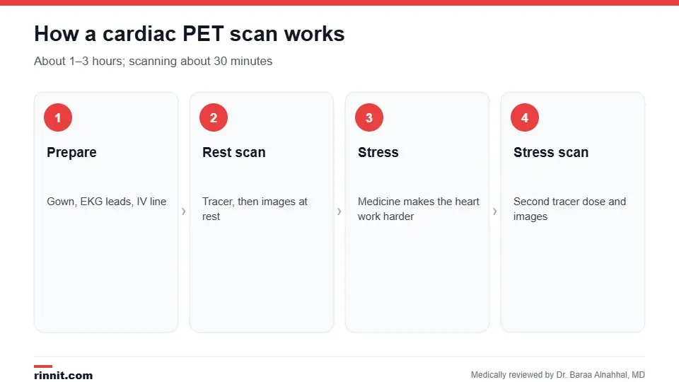
Rest and stress images are compared.
You change into a gown, have your blood pressure checked and get EKG leads and an IV.
Rest phase: a small tracer dose is given and images are taken.
Stress phase: medicine through the IV makes your heart respond as if you were exercising; you may feel warm, cold or a mild headache.
A second tracer dose is given and stress images are taken.
The IV is removed, and you drink plenty of water for 24 hours. Sources: [1]
Tell the staff right away about chest pain, arm or jaw discomfort, shortness of breath or dizziness. Sources: [1]
Risks
Radiation exposure is low. Redness or soreness at the IV site is common; an allergic reaction to the tracer is rare. Tell your provider if you're pregnant or breastfeeding. Diabetes can make results less accurate, and some people with a BMI over 30 may need a special scanner. A sedative can help with claustrophobia. Sources: [1]
Understanding your results
Results usually reach your provider within 24–48 hours. They show which areas of heart muscle get enough blood and which may improve with treatment. Abnormal results may lead to angioplasty, stents, bypass surgery or medicine changes. If damage is too extensive for a procedure to help, continued medicine may be recommended. Sources: [1, 2, 3]
Conclusion
A cardiac PET scan is one of the most accurate noninvasive ways to check blood flow to the heart. It helps your care team decide whether you need a procedure and how well treatment is working.
Keep learning: read about coronary artery disease and the nuclear stress test.
Frequently asked questions
What is a cardiac PET scan?
A nuclear imaging test showing heart blood flow and muscle health.
Why is it done?
To diagnose coronary artery disease and check heart damage.
How long does it take?
About 1–3 hours, with about 30 minutes of scanning.
Do I need to fast?
Yes, nothing but water for 4 hours.
Can I have coffee?
No caffeine for 24 hours, including decaf.
Will I have to exercise?
No. Medicine makes your heart work as if exercising.
Is it safe?
Yes. Radiation is low and serious side effects are rare.
Is the tracer a dye?
No. It's a small radioactive tracer, not a contrast dye.
When will I get results?
Usually within 24–48 hours.
How is PET different from SPECT?
PET generally gives sharper images and higher accuracy.
Related Reading
Coronary Flow Reserve (CFR): What It Measures and Normal Values explains how coronary flow reserve shows the heart's ability to boost blood flow.
Coronary Artery Disease: Symptoms, Causes, Diagnosis, Treatment and Prevention — What You Need to Know the main condition cardiac PET evaluates.
Nuclear Stress Test: How It Works, How to Prepare and What Results Mean a related nuclear heart test.
PET Scan: Uses, Preparation, What to Expect and Results how PET imaging works.
Cardiac Sarcoidosis: When Immune Cell Clumps Affect Your Heart — Symptoms, Tests, and Treatment inflammation PET can detect.
SPECT Scan: How It Works, What It Shows and What to Expect another nuclear imaging method.
References
Cleveland Clinic. Heart PET Scan. Cleveland Clinic. Last updated April 25, 2023. https://my.clevelandclinic.org/health/diagnostics/17376-cardiac-positron-emission-tomography-pet
Parker MW, Iskandar A, Limone B, et al. Diagnostic accuracy of cardiac positron emission tomography versus single photon emission computed tomography for coronary artery disease: a bivariate meta-analysis. Circ Cardiovasc Imaging. 2012;5(6):700-707. PMID 23051888. https://doi.org/10.1161/CIRCIMAGING.112.978270
Danad I, Raijmakers PG, Driessen RS, et al. Comparison of coronary CT angiography, SPECT, PET, and hybrid imaging for diagnosis of ischemic heart disease determined by fractional flow reserve. JAMA Cardiol. 2017;2(10):1100-1107. PMID 28813561. https://doi.org/10.1001/jamacardio.2017.2471
Medical disclaimer
This article is for general education only and does not replace advice from a qualified healthcare professional. Talk to your doctor about your own diagnosis and treatment, and seek emergency care right away for severe or sudden symptoms.



Comments