Heart MRI (Cardiac MRI): What It Shows, How to Prepare, What Happens and Results

Updated: 2 days ago
Written and medically reviewed by Dr. Baraa Alnahhal, MD · Last reviewed: November 2026 · How we review
Quick Answer: What is a heart MRI?
A heart MRI, or cardiac MRI, is a noninvasive scan that uses a strong magnet and radio waves, not radiation, to make detailed pictures of the heart. It shows the heart's chambers, valves and muscle and how well they work. It takes 30 minutes to 1.5 hours. [1]
What the research says
Heart MRI picks up blocked arteries better than a stress echo. In 47 studies with 4,742 people, a stress perfusion heart MRI found significant coronary narrowing with 88% sensitivity, compared with 72% for dobutamine stress echocardiography, with similar specificity. (Haberkorn et al., Frontiers in Cardiovascular Medicine, 2021) [2]
Scarring seen on heart MRI helps predict outlook in myocarditis. In 8 studies of 1,319 people with myocarditis, those with scar-like late gadolinium enhancement had about 4.6 times higher odds of a major heart event. (Yang et al., European Radiology, 2020) [3]
Heart MRI often changes the diagnosis after an unexplained heart attack. In 26 studies of 3,624 people with a heart attack but no blocked arteries, 68% were given a different diagnosis after MRI, most often myocarditis (31%). (Mileva et al., JACC: Cardiovascular Imaging, 2023) [4]
Key takeaways
A heart MRI uses a magnet and radio waves to create detailed pictures of your heart, with no radiation. [1]
It shows both the heart's structure and how well it pumps and moves blood. [1]
Contrast dye given through an IV may be used to highlight problem areas. [1]
You will need to hold your breath for parts of the scan, which takes 30 minutes to 1.5 hours. [1]
Metal in your body and some implanted devices can make the scan unsafe, so tell your provider about them. [1]
Results usually come back within a few days. [1]
What is a heart MRI?
A heart MRI is an imaging test that gives your provider a detailed look at your heart without surgery or radiation. Nothing is placed inside your body that you can see or feel. [1]
The scanner uses a large magnet and radio waves to pick up tiny changes in the molecules in your body. A computer turns those signals into 2D or 3D images of your heart's chambers, valves and muscle. The images also show how well each part works and how blood moves through your heart. [1]
Sometimes a contrast dye is given through a vein to make problem areas stand out more clearly. [1]
How is a heart MRI different from a regular MRI?
The main difference is that you hold your breath at several points during a heart MRI. This keeps your chest still so the moving heart can be captured clearly. [1]
Why is a heart MRI done?

A heart MRI shows both the structure and the function of your heart.
Your provider may order a heart MRI to study the structure and function of your heart. Common reasons include: [1]
Finding the cause of chest pain, shortness of breath or fainting
Diagnosing congenital heart disease, heart failure, heart valve disease, a mass in the heart or inflammation of the sac around the heart
Looking for heart muscle damage, infection or inflammation
Detecting buildup of protein (amyloidosis) or iron (hemochromatosis) in the heart muscle
Checking damage after a heart attack or areas with poor blood flow from blocked arteries
Finding the exact spots to target during an ablation procedure
How else is a heart MRI used?
Beyond diagnosis, providers use it to see how well a treatment is working, plan procedures, check the results of heart surgery and follow how a condition changes over time. [1]
How accurate is a heart MRI?
A heart MRI is very accurate. It often finds disease better than a CT scan, and it can show how severe valve disease is and how much heart tissue has been damaged. [1]
What the scan looks at | What it can reveal |
Heart chambers and muscle | Size, thickness, pumping strength and muscle damage |
Heart valves | Valve disease and how severe it is |
Blood flow | Areas with poor blood supply from blocked arteries |
Heart tissue | Inflammation, infection, scarring, protein or iron buildup |
Sac around the heart | Inflammation of the pericardium |
Sources: [1]
How do you prepare for a heart MRI?
Tell your provider about any allergies, health problems and past surgeries. Your kidney function also matters, because your kidneys have to clear the contrast dye from your body. If your kidneys aren't working well, your provider may limit the dye or skip it. [1]
What if you are anxious or claustrophobic?
If small spaces make you nervous, ask your provider for a sedative a few days before the scan. You pick it up at a pharmacy and take it about 30 minutes before your appointment. You will need someone to drive you home afterward. Open MRI machines, which aren't fully enclosed, are another option if you need more space. [1]
What should you avoid on the day of the scan?
On the day of your scan, plan to: [1]
Leave jewelry and valuables at home
Remove metal items such as piercings, glasses, hearing aids and hair clips, and store them in a locker
Avoid clothes with metal, such as zippers or underwire; you may change into a gown, but you can usually keep on underwear and socks
Keep taking your usual medicines and eating normally unless you're told otherwise
Empty your bladder before the scan
What happens during a heart MRI?
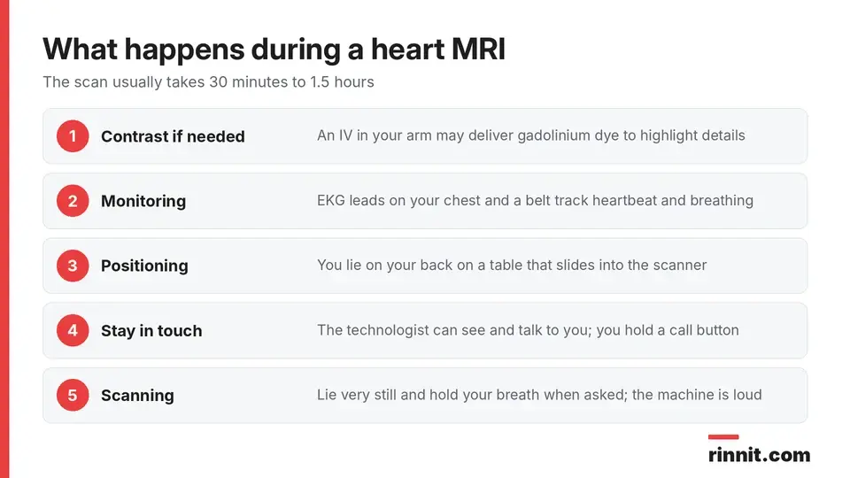
A heart MRI usually takes 30 minutes to 1.5 hours.
The MRI machine looks like a large doughnut standing on its edge. The scan generally follows these steps: [1]
Contrast (if needed): A technologist may place an IV in your arm to give gadolinium contrast dye.
Monitoring: EKG leads go on your chest, and a belt around your lower chest tracks your heartbeat and breathing.
Positioning: You lie on your back on a table that slides into the scanner.
Staying in touch: The technologist sits behind a glass window but can see and talk to you through an intercom. You hold a call button and may get earplugs or music.
Scanning: You lie very still while the machine takes a series of scans, each lasting a few minutes. You will be asked to hold your breath at times.
The machine is very loud. Lying still is important, because any movement can blur the pictures. [1]
How long does a heart MRI take?
The whole test takes about 30 minutes to 1.5 hours, depending on how many scans your provider needs. [1]
What happens after a heart MRI?
The table slides out of the scanner, and the IV is removed if you had one. You can get dressed and go home. Drinking plenty of water helps your body flush out the contrast dye. If you took a sedative, you may feel drowsy until it wears off. [1]
What are the risks of a heart MRI?
A heart MRI is generally safe and does not expose you to radiation. Your provider will ask whether you are pregnant and whether you have any metal in your body, because the test isn't right for everyone. [1]
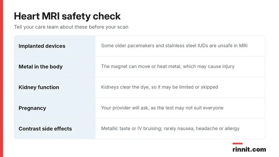
Tell your care team about metal, devices and kidney problems before the scan.
Risk factor | Why it matters |
Implanted devices | Some, such as certain older pacemakers and stainless steel IUDs, are unsafe or can interfere with the scan |
Metal in the body | The magnet can move metal or make it heat up, which may cause injury |
Contrast dye | Can cause a metallic taste or bruising at the IV site, and rarely nausea, headache or an allergic reaction |
Sedation | You may feel drowsy until it wears off |
Kidney problems | Your kidneys must clear the dye, so your provider may limit or avoid it |
Sources: [1]
Your provider can check a medical device safety database to see whether your implant is safe in an MRI. [1]
What do heart MRI results mean?
A radiologist or cardiologist studies your images and sends a report to the provider who ordered the test. Results usually take a few days, and your provider may go over the images with you at your next visit. [1]
If the results are abnormal, your next steps depend on the condition found and may include medicines, a procedure or surgery. [1]
When should you call your provider?
Get medical help right away if you have heart symptoms such as chest pain. If you haven't heard about your results after a few days, contact your provider's office to follow up. [1]
Conclusion
A heart MRI gives your provider one of the most detailed views of your heart's structure and function, without radiation. It helps explain symptoms, diagnose many heart conditions and guide treatment.
Before your scan, tell your care team about any metal, implanted devices, kidney problems or pregnancy, and ask about sedation if tight spaces worry you.
Keep learning: Read our MRI guide or our echocardiogram guide. For more background, see RadiologyInfo on cardiac MRI.
FAQ
What is a heart MRI?
It is a scan that uses a magnet and radio waves to make detailed pictures of your heart's structure and function.
Does a heart MRI use radiation?
No. It uses magnets and radio waves instead of radiation.
How long does a cardiac MRI take?
It usually takes about 30 minutes to 1.5 hours.
What can a heart MRI detect?
It can show heart failure, valve disease, congenital heart disease, inflammation, heart muscle damage and protein or iron buildup.
Why do I have to hold my breath during a heart MRI?
Holding your breath keeps your chest still so the images of your moving heart stay sharp.
Is contrast dye always used for a heart MRI?
No. Gadolinium dye is used only when your provider needs extra detail.
Can I have a heart MRI with a pacemaker?
It depends on the device; some older pacemakers are unsafe, so your provider checks your device first.
What if I'm claustrophobic?
You can ask for a sedative ahead of time or ask whether an open MRI machine is available.
Is a heart MRI better than a CT scan?
A heart MRI often finds heart disease better than CT and can show how much tissue is damaged.
When will I get my heart MRI results?
Results usually come back within a few days.
Related Reading
MUGA Scan: Measuring How Well Your Heart Pumps explains the nuclear scan that measures ejection fraction.
Heart Biopsy (Endomyocardial Biopsy): Procedure, Risks and Results explains the catheter procedure that takes tiny samples of heart muscle.
Intraoperative MRI (iMRI): MRI Scans During Brain Surgery explains MRI scanning during surgery to check tumor removal.
Cardiac MRI Adenosine Stress Test: Prep, Side Effects and Results explains the stress heart MRI that uses adenosine instead of exercise.
Cardiac Imaging: Types of Heart Scans and What Each One Shows compares the main heart imaging tests and what each one shows.
What Is Myocarditis? A Plain-Language Guide to the Heart Muscle Inflammation That Often Hides explains a heart muscle inflammation that a heart MRI can reveal.
Congenital Heart Disease (CHD): Types, Symptoms, Treatment & Survival — Complete Guide covers heart defects present at birth that a heart MRI can map in detail.
ATTR-CM (Transthyretin Amyloid Cardiomyopathy): The Rare Heart Condition Caused by Protein Clumps — Explained in Plain English describes protein buildup in the heart muscle that a heart MRI can detect.
Understanding Restrictive Cardiomyopathy: Causes, Symptoms, and Heart Health explains a stiff heart muscle condition where a heart MRI helps find the cause.
Heart Failure: Symptoms, Causes & Treatment Guide covers a condition a heart MRI can help diagnose and monitor.
Constrictive Pericarditis: What It Is, Symptoms, Causes, and Treatment describes inflammation of the sac around the heart that a heart MRI can show.
References
Cleveland Clinic. Cardiac MRI: Procedure Details & Uses. Cleveland Clinic. Last updated January 7, 2025. https://my.clevelandclinic.org/health/diagnostics/21961-heart-mri
Haberkorn SM, Haberkorn SI, Bönner F, et al. Vasodilator Myocardial Perfusion Cardiac Magnetic Resonance Imaging Is Superior to Dobutamine Stress Echocardiography in the Detection of Relevant Coronary Artery Stenosis: A Systematic Review and Meta-Analysis on Their Diagnostic Accuracy. Front Cardiovasc Med. 2021;8:630846. PMID 33778024. https://doi.org/10.3389/fcvm.2021.630846
Yang F, Wang J, Li W, et al. The prognostic value of late gadolinium enhancement in myocarditis and clinically suspected myocarditis: systematic review and meta-analysis. Eur Radiol. 2020;30(5):2616-2626. PMID 32040731. https://doi.org/10.1007/s00330-019-06643-5
Mileva N, Paolisso P, Gallinoro E, et al. Diagnostic and Prognostic Role of Cardiac Magnetic Resonance in MINOCA: Systematic Review and Meta-Analysis. JACC Cardiovasc Imaging. 2023;16(3):376-389. PMID 36889851. https://doi.org/10.1016/j.jcmg.2022.12.029
Medical disclaimer
This article is for general education only and does not replace advice from a qualified healthcare professional. Talk to your doctor about your own diagnosis and treatment, and seek emergency care right away for severe or sudden symptoms.



Comments