Intraoperative MRI (iMRI): MRI Scans During Brain Surgery

Updated: 2 days ago
Written and medically reviewed by Dr. Baraa Alnahhal, MD · Last reviewed: November 2026 · How we review
Quick Answer
Intraoperative MRI (iMRI) is an MRI scan done during surgery that gives the surgical team a clear, up-to-date view of the area they're operating on. It's used most often in brain surgery, such as removing brain tumors. It helps surgeons confirm the whole tumor has been removed before closing, which can reduce the need for a second surgery.
What the research says
iMRI leads to more complete tumor removal: in a randomized trial of 58 people having glioma surgery, the whole visible tumor was removed in 96% with intraoperative MRI vs. 68% without it (Senft C et al., Lancet Oncology, 2011) [2]
Reviews support the benefit: a Cochrane review found intraoperative MRI reduced the chance of incomplete glioma removal, though the evidence came from small trials and was rated very low quality (Jenkinson MD et al., Cochrane Database of Systematic Reviews, 2018) [3]
Key takeaways
Intraoperative MRI takes MRI pictures during surgery.
It helps surgeons check that a tumor has been fully removed.
It's mainly used in brain, pituitary and epilepsy surgery.
Images are available within seconds to minutes.
It uses no radiation, but metal in the body must be checked first.
What is intraoperative MRI?
During brain surgery, the brain can shift slightly once the skull is opened, so scans taken beforehand may no longer match exactly. With iMRI, the team can scan during the operation to see what remains. MRI uses strong magnets and radio waves: the magnet briefly aligns protons in your tissues, radio pulses disturb them, and as they realign the scanner records signals that a computer turns into images. Sources: [1]
Why it's used
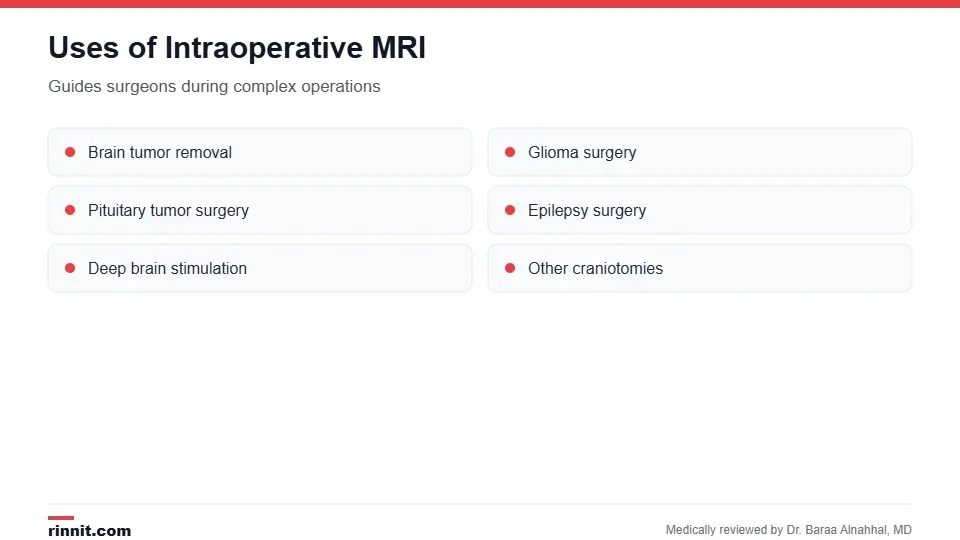
iMRI guides surgeons during complex operations.
Removing brain tumors, including gliomas.
Pituitary tumor surgery.
Epilepsy surgery.
Placing deep brain stimulation devices for Parkinson's disease.
Other craniotomies. Sources: [1]
Benefits and limits

Your surgeon decides whether iMRI suits your operation.
Benefits | Limits |
Confirms how much tumor was removed | The scanner is loud |
May reduce the need for a second surgery | Metal in or on the body can be a safety hazard |
Images ready in seconds to minutes | Not available at every hospital |
No radiation | Adds time to the operation |
Sources: [1, 2, 3]
How to prepare
There's no special preparation for iMRI itself; follow your surgeon's instructions.
Tell your care team if you're pregnant or have metal in your body, such as a pacemaker or implants.
Remove jewelry and other metal, such as earrings, hairpins and underwire bras. Sources: [1]
What happens during surgery

The scan fits into the operation without long delays.
You receive general anesthesia; some procedures are done awake while the team keeps you comfortable.
The surgeon begins the operation.
At key points, an MRI scan is taken in the operating room.
The surgeon reviews the images within seconds to minutes.
If tumor remains and it's safe to remove, the surgeon continues before closing. Sources: [1]
Risks
iMRI is painless and uses no radiation. The main safety concern is metal, which can move or malfunction in the magnetic field, so careful screening is done beforehand. Risks of the surgery itself are separate and depend on the operation. Sources: [1]
Conclusion
Intraoperative MRI lets surgeons check their work in real time, especially when removing brain tumors. Seeing what remains before closing can mean a more complete operation and a lower chance of needing another surgery.
Keep learning: read about brain tumors and MRA.
Frequently asked questions
What is intraoperative MRI?
An MRI scan done during surgery.
Why is iMRI used?
To help surgeons confirm a tumor has been fully removed.
What surgeries use iMRI?
Mainly brain tumor, pituitary, epilepsy and deep brain stimulation surgery.
Will I be awake?
Usually no; most people have general anesthesia.
Does iMRI use radiation?
No.
How fast are the images ready?
Within seconds to minutes.
Can it prevent a second surgery?
It may lower the chance of needing one.
Is metal a problem?
Yes. Tell your team about any implants or devices.
Is the scan painful?
No, but the scanner is loud.
Is iMRI available everywhere?
No. Only some hospitals have it.
Related Reading
Brain Tumor: Symptoms, Causes, Diagnosis and Treatment — What You Need to Know a main reason for iMRI-guided surgery.
MRA (Magnetic Resonance Angiography): What It Is, Why It's Done, Procedure and Results MRI imaging of blood vessels.
Electroencephalogram (EEG): Types, How to Prepare and What Results Mean a test used to plan epilepsy surgery.
Gadolinium Contrast for MRI: How It Works, Side Effects and Kidney Safety the contrast often used in MRI.
Heart MRI (Cardiac MRI): What It Shows, How to Prepare, What Happens and Results another use of MRI imaging.
References
Cleveland Clinic. Intraoperative MRI (iMRI): What It Is, Purpose & Advantages. Cleveland Clinic. Last updated January 18, 2024. https://my.clevelandclinic.org/health/diagnostics/intraoperative-mri
Senft C, Bink A, Franz K, et al. Intraoperative MRI guidance and extent of resection in glioma surgery: a randomised, controlled trial. Lancet Oncology. 2011;12(11):997-1003. PMID 21868284. https://doi.org/10.1016/S1470-2045(11)70196-6
Jenkinson MD, Barone DG, Bryant A, et al. Intraoperative imaging technology to maximise extent of resection for glioma. Cochrane Database of Systematic Reviews. 2018;1(1):CD012788. PMID 29355914. https://doi.org/10.1002/14651858.CD012788.pub2
Medical disclaimer
This article is for general education only and does not replace advice from a qualified healthcare professional. Talk to your doctor about your own diagnosis and treatment, and seek emergency care right away for severe or sudden symptoms.



Comments