MRA (Magnetic Resonance Angiography): What It Is, Why It's Done, Procedure and Results

Updated: 2 days ago
Written and medically reviewed by Dr. Baraa Alnahhal, MD · Last reviewed: November 2026 · How we review
Quick Answer: What is an MRA?
An MRA (magnetic resonance angiography) is a type of MRI scan that creates detailed pictures of the arteries, veins and the blood flowing through them. A provider may order it to look for narrowed, widened or blocked blood vessels, such as an aneurysm or a buildup of plaque. It takes 20 to 60 minutes and uses no radiation.
What the research says
MRA performs about as well as CT angiography for finding brain aneurysms. A meta-analysis of 10 studies found that MRA had a pooled sensitivity of 80% and specificity of 87% for intracranial aneurysms, with no significant difference in overall accuracy compared with CT angiography. (Chen et al., Medicine, 2018) [2]
Contrast-enhanced MRA is highly accurate for leg artery blockages. Across 32 prospective studies with 1,022 people who had symptoms of peripheral artery disease, MRA had a pooled sensitivity of 94.7% and specificity of 95.6% for narrowings of 50% or more, and it correctly classified 95.3% of artery segments. (Menke and Larsen, Annals of Internal Medicine, 2010) [3]
MRA reliably detects severe narrowing and blockage of the carotid artery. In a meta-analysis, contrast-enhanced MRA found 70% to 99% internal carotid stenosis with 94.6% sensitivity and 91.9% specificity, and it detected full blockages with 99.4% sensitivity. Its sensitivity for moderate (50% to 69%) narrowing was lower, at 65.9%. (Debrey et al., Stroke, 2008) [4]
Key takeaways
An MRA is a form of MRI that focuses on your blood vessels and how blood moves through them [1].
It can find narrowed, bulging or blocked vessels without placing a catheter inside your body [1].
The test uses a strong magnet and radio waves, not X-ray radiation [1].
Some MRAs use contrast dye through an IV, while others are done without any dye [1].
The scan usually takes 20 to 60 minutes, and you can often go back to normal activities right away [1].
Results typically take a few days, and a report that reads "unremarkable" means nothing concerning was seen [1].
What is an MRA?

An MRA builds pictures of your blood vessels using magnetism and radio waves.
An MRA, or magnetic resonance angiography, is an imaging test that shows your blood vessels and the flow of blood inside them. It is a specialized type of MRI that centers on your arteries and veins [1].
The scan can reveal whether a blood vessel has become narrow, enlarged or blocked [1].
Traditional angiography requires a thin tube called a catheter to be threaded into a blood vessel. An MRA can gather similar information without that step, which makes it noninvasive [1].
How the scan works
The machine uses a powerful magnetic field and radio waves to create images. It does not use the ionizing radiation found in X-rays or CT scans [1].
The images appear on a computer, where a specialist can study your vessels from different angles [1].
Why is an MRA done?
Your provider may suggest an MRA when they need a clear look at blood vessels in a specific part of your body. It can confirm a suspected condition and show how serious it is [1].
Body areas an MRA can image
The aorta, the largest artery in your body [1].
Arteries throughout the body, for example when vasculitis or atherosclerosis is suspected [1].
Blood vessels in the brain [1].
The carotid arteries in your neck [1].
The coronary arteries that supply your heart [1].
Peripheral arteries, such as those in your legs [1].
The pulmonary arteries in your lungs [1].
The renal arteries that supply your kidneys [1].
Conditions an MRA can help find
Condition | What it means |
Aneurysm | A weakened, bulging area in a blood vessel wall |
Aortic dissection | A tear in the inner layer of the aorta |
Arteriovenous malformation (AVM) | An abnormal tangle of arteries and veins |
Artery stenosis | Narrowing of an artery |
Atherosclerosis | Plaque buildup inside artery walls |
Congenital heart disease | Heart or vessel differences present from birth |
Deep vein thrombosis (DVT) | A blood clot in a deep vein, often in the leg |
Pulmonary embolism | A blood clot that travels to the lungs |
Sources: [1]
Brain MRA
A brain MRA can detect brain aneurysms, signs of a stroke and vascular malformations [1].
Your provider may also use it to help evaluate secondary headaches, which are headaches caused by another health problem [1].
Types of MRA
Your care team chooses the type of MRA based on the vessels they need to see and your overall health [1].
Type | How it differs | When it is typically used |
Contrast-enhanced MRA | Contrast dye is given through an IV | To make blood vessels easier to see |
Non-enhanced MRA | No contrast dye is used | When contrast is not safe for you |
2D MRA | Captures flat image slices | Long stretches of a blood vessel |
3D MRA | Captures a full volume of tissue | Compact groups of arteries, such as the circle of Willis in the brain |
Sources: [1]
How do you prepare for an MRA?
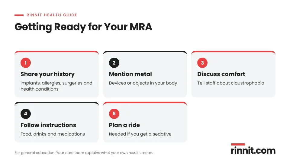
Sharing your health history ahead of time helps your MRA go safely and smoothly.
Because the scanner uses a very strong magnet, your care team needs to know about anything that could interact with it or affect your safety [1].
What to tell your provider
Tell your provider about | Why it matters |
Metal in your body | Medical devices or foreign objects can be affected by the magnet |
Claustrophobia | The scanner is an enclosed space, and options can help you feel calmer |
Pregnancy or breastfeeding | Your team may adjust how the test is done |
Health conditions, allergies and past surgeries | These help your team plan a safe scan |
Kidney or liver disease | These conditions may mean you cannot receive contrast dye |
Sources: [1]
Before the day of the test
What happens during an MRA?

Most MRA scans take between 20 and 60 minutes from start to finish.
You lie on a padded table that slides into a large scanner shaped like a donut. The machine is loud, so you will get headphones or earplugs [1].
It is important to stay very still so the pictures are clear. At times, you may be asked to hold your breath for a short period [1].
The technologist works from a nearby room and can see you throughout the scan. You will hold a small ball that you can squeeze if you need their attention [1].
If you receive contrast dye
When your scan uses contrast, the dye goes in through an IV line. Some people notice a brief metallic taste as it enters the bloodstream [1].
How long it takes
An MRA usually lasts 20 to 60 minutes [1].
Does an MRA hurt?
The scan itself does not cause pain. The only physical sensation for most people is the IV placement when contrast is used [1].
If noise or the enclosed space worries you, headphones and a sedative can help you stay comfortable [1].
What are the risks of an MRA?
An MRA is considered safe and noninvasive. It does not expose you to radiation [1].
The only substance that may enter your body is the contrast dye. If kidney failure is a concern, the test can be done without dye [1].
Side effects from contrast are uncommon. They can include a headache, an upset stomach or an allergic reaction [1].
What happens after an MRA?
If you did not receive a sedative, you can usually drive yourself and return to your regular activities right away [1].
If you were sedated, someone else will need to drive you home [1].
What do MRA results mean?
A radiologist, a doctor who specializes in reading medical images, reviews your scan. It usually takes a few days for them to send the results to your provider [1].
If your report says "unremarkable," it means the radiologist did not see anything of concern [1].
When a problem is found, the MRA can help confirm the diagnosis and show how severe it is. Your provider will explain what the findings mean for you and discuss next steps [1].
When to contact your provider
If you have not heard about your results after a week, reach out to your provider's office [1].
Conclusion
An MRA gives your care team a detailed view of your blood vessels without a catheter or radiation. It can help find problems such as aneurysms, narrowed arteries and blood clots, and it can show how serious they are.
Preparing well, by sharing your health history and following your team's instructions, helps the scan go smoothly. If you have questions about your results, your provider is the best person to talk with.
Keep learning: Read our complete guide to MRI scans or our guide to how an angiogram works. For more background, see the MedlinePlus MR angiography page.
FAQ
What is an MRA?
An MRA is a type of MRI scan that makes detailed pictures of your blood vessels and shows how blood flows through them.
Why would my doctor order an MRA?
It helps find narrowed, widened or blocked blood vessels, such as aneurysms, plaque buildup, blood clots or vessel malformations.
How is an MRA different from a regular MRI?
Both use the same type of scanner, but an MRA is set up to focus on arteries and veins rather than other tissues and organs.
Does an MRA use radiation?
No. An MRA uses a strong magnetic field and radio waves, so you are not exposed to X-ray radiation.
Does an MRA hurt?
The scan is painless. If contrast dye is used, you may feel a small pinch when the IV is placed.
How do I prepare for an MRA?
Tell your provider about any metal in your body, allergies, pregnancy, kidney or liver disease, and follow their food and medication instructions.
How long does an MRA take?
Most MRA scans take between 20 and 60 minutes.
Is contrast dye always used for an MRA?
No. Some MRAs use dye to make vessels clearer, while non-enhanced MRAs are done without it when dye is not safe for you.
What are the risks of an MRA?
An MRA is very safe. Rare side effects from contrast dye include headache, upset stomach or an allergic reaction.
What does an unremarkable MRA result mean?
It means the radiologist did not see anything concerning on your scan.
How soon will I get my MRA results?
Results usually reach your provider within a few days. Contact your provider if you have not heard back after a week.
Related Reading
Intraoperative MRI (iMRI): MRI Scans During Brain Surgery explains MRI scanning during surgery to check tumor removal.
Brain Aneurysm: Symptoms, Causes, and Treatment explains the bulging brain vessels that a brain MRA is often used to find.
Stroke: Symptoms, Causes, Diagnosis, and Treatment — A Complete Guide covers how imaging such as MRA helps show signs of stroke and its causes.
Carotid Artery Disease: The Silent Stroke Risk in Your Neck, Explained explains the neck artery narrowing that an MRA can measure.
Peripheral Artery Disease (PAD): Symptoms, Causes, Diagnosis & Treatment Options covers the leg artery blockages that contrast-enhanced MRA can detect.
Narrowed Kidney Arteries: What Renal Artery Stenosis Is, Why It Happens, and How It Is Treated explains a kidney artery problem that an MRA of the renal arteries can reveal.
Brain AVM (Arteriovenous Malformation): Symptoms, Causes, and Treatment covers the abnormal vessel tangles that a brain MRA can help identify.
Aortic Aneurysm: Types, Causes, Dissection Risk & Complete Guide explains aortic aneurysms and dissections, both of which an MRA of the aorta can show.
References
Cleveland Clinic. Magnetic Resonance Angiography (MRA). Cleveland Clinic. Last updated April 24, 2025. https://my.clevelandclinic.org/health/diagnostics/24024-mra
Chen X, Liu Y, Tong H, et al. Meta-analysis of computed tomography angiography versus magnetic resonance angiography for intracranial aneurysm. Medicine (Baltimore). 2018;97(20):e10771. PMID 29768368. https://doi.org/10.1097/MD.0000000000010771
Menke J, Larsen J. Meta-analysis: Accuracy of contrast-enhanced magnetic resonance angiography for assessing steno-occlusions in peripheral arterial disease. Ann Intern Med. 2010;153(5):325-34. PMID 20820041. https://doi.org/10.7326/0003-4819-153-5-201009070-00007
Debrey SM, Yu H, Lynch JK, et al. Diagnostic accuracy of magnetic resonance angiography for internal carotid artery disease: a systematic review and meta-analysis. Stroke. 2008;39(8):2237-48. PMID 18556586. https://doi.org/10.1161/STROKEAHA.107.509877
Medical disclaimer
This article is for general education only and does not replace advice from a qualified healthcare professional. Talk to your doctor about your own diagnosis and treatment, and seek emergency care right away for severe or sudden symptoms.



Comments