MRI (Magnetic Resonance Imaging): Uses, What to Expect, Safety and Results

Updated: 6 days ago
Written and medically reviewed by Dr. Baraa Alnahhal, MD · Last reviewed: November 2026 · How we review
Quick Answer: What is MRI (magnetic resonance imaging)?
An MRI is a painless imaging test that uses a large magnet, radio waves and a computer to create detailed pictures of soft tissues such as the brain, nerves, muscles, ligaments and organs. It uses no radiation, takes about 30 to 60 minutes, and is low risk when safety screening for metal and implants is followed [1].
What the research says
Gadolinium contrast reactions are uncommon in children. A meta-analysis covering 112,760 contrast-enhanced MRI exams found acute reaction rates of 0.25% for physiologic and 0.13% for allergic-like reactions, delayed reactions in 0.04%, and no cases of nephrogenic systemic fibrosis among 4,895 patients. (Yu et al., Radiology, 2025) [2]
Contrast MRI was not linked to more kidney injury in adults. In a propensity-matched study of 35,197 inpatient MRI exams, acute kidney injury occurred in 1.62% overall and was less common after gadolinium contrast (odds ratio 0.59), including in people with moderately reduced kidney function. (Han et al., Investigative Radiology, 2026) [3]
Preparation and distraction reduce the need for sedation in kids. Across 20 studies with 33,873 children and adolescents, non-drug strategies were linked to less sedation or general anesthesia for MRI (risk ratio 0.68 in randomized trials), with the biggest effect in children aged 3 to 10. (Thestrup et al., Hospital Pediatrics, 2023) [4]
Key takeaways
MRI makes detailed images of soft tissue using a magnet and radio waves, not ionizing radiation [1].
It helps evaluate the brain, spine, heart, abdominal and pelvic organs, breasts, bones, joints and blood vessels [1].
The magnet can affect implanted devices and metal, so you must tell your care team about any implant or metal fragment before the scan [1].
Most scans take 30 to 60 minutes, are painless and loud, and require you to lie very still [1].
Contrast dye is sometimes injected to highlight tumors, inflammation and blood vessels; serious reactions are rare [1, 2].
A radiologist reads the images, and results usually reach your provider within a few days [1].
What is an MRI scan?
Magnetic resonance imaging (MRI) is a diagnostic imaging test that produces detailed pictures of the inside of your body. It is especially good at showing soft tissues, such as muscles, ligaments, nerves and organs, that can be hard to see on other tests [1].
A traditional MRI scanner is a large, tunnel-shaped machine. You lie on a padded table that slides into the opening while the scanner collects images [1].
Unlike X-rays and CT scans, MRI does not use radiation. The magnetic field is not harmful itself, but it can affect metal objects and implanted devices, so safety screening comes first [1].
What is an MRI used for?
The scan can examine the brain and nerve tissue, the spine and spinal cord, the heart and major blood vessels, chest and abdominal organs such as the liver, kidneys, spleen, pancreas, bowel and adrenal glands, pelvic organs such as the bladder and reproductive organs, breast tissue, and bones and joints [1].
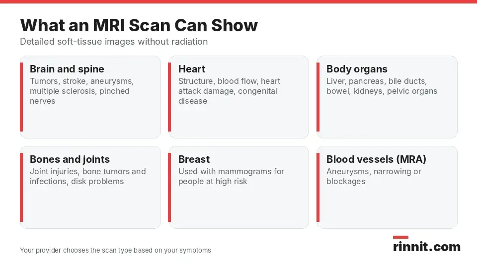
Each type of MRI focuses on a different body region, and your provider chooses the one that matches your symptoms.
MRI type | What it can help evaluate |
Brain and spine MRI | Brain and spinal tumors, brain aneurysms, stroke, multiple sclerosis, spinal cord injuries and pinched nerves |
Functional MRI (fMRI) | Brain activity, measured through changes in blood flow; often used to map the brain before surgery |
Cardiac (heart) MRI | Heart structure and function, blood flow, damage from coronary artery disease or heart attack, congenital heart disease, and heart tumors, infections or inflammation |
Body MRI | Tumors in the chest, abdomen or pelvis; liver, pancreas and bile duct conditions; inflammatory bowel disease; blood vessel problems and inflammation |
Bone and joint MRI | Bone tumors and infections, joint injuries, and spine and disk problems |
Breast MRI | Used along with mammograms to help find breast cancer in people at high risk |
Magnetic resonance angiography (MRA) | Blood vessels, to find aneurysms, narrowing or blockages |
Sources: [1]
How does an MRI work?
Electric current running through coils of wire inside the scanner creates a strong, temporary magnetic field. The scanner then sends radio waves into your body [1].
These radio waves cause certain atoms in your tissues to give off signals. A receiver picks up those signals, and a computer turns them into detailed digital images that a radiologist can view from many angles [1].
What are the different types of MRI?
What is an open MRI?
An open MRI uses a machine with a wider, open-sided design instead of a narrow tunnel. It can be a helpful option if you have claustrophobia. Some facilities also offer upright scanners that let you sit, stand or bend during imaging [1].
What is an MRI with contrast?
For some scans, a contrast dye is injected through an IV line in your arm or hand. Contrast makes tumors, inflammation, blood vessels and blood flow easier to see [1].
Contrast is generally safe, and serious reactions are rare [1, 2]. If you are pregnant or might be, or if you have kidney disease, tell your provider before receiving contrast; in severe kidney disease, gadolinium can rarely cause a serious condition called nephrogenic systemic fibrosis [1].
How do I prepare for an MRI?
Preparation focuses on safety. Because the magnet is always on, your care team needs to know about anything in or on your body that contains metal or electronics [1].
What implants and devices should I tell my care team about?
Tell your provider and the MRI technologist if you have any of the following [1]:
A heart pacemaker or defibrillator
An electronic or implanted stimulator or other implanted device
A metal joint replacement
A cochlear implant or other ear implant
An implanted drug pump
A programmable shunt
Aneurysm clips or coils
A stent outside the heart
A filter, such as a blood clot filter
Metal fragments in your body or eye, such as bullets, shrapnel or metal shavings
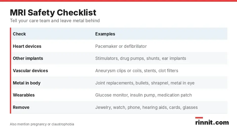
Reviewing this checklist with your care team before the scan day helps avoid delays and keeps the exam safe.
Some wearable devices cannot stay on during the scan. If you use a continuous glucose monitor, an insulin pump or a medication patch, ask how to schedule your MRI around it [1].
What else should I tell my provider?
Let your provider know ahead of time if you [1]:
Are pregnant or think you could be
Cannot lie flat on your back for 30 to 60 minutes
Have claustrophobia or feel anxious in tight spaces
What should I remove before the scan?
You will need to take off or leave behind items that contain metal or electronics [1].
Category | Items to remove or leave at home |
Jewelry and accessories | Jewelry, watches, body piercings, hair pins and metal hair accessories |
Clothing | Underwire bras and clothing with metal zippers |
Personal devices | Phones, electronic tracking devices and hearing aids |
Everyday items | Credit cards, pens, pocketknives and eyeglasses |
Dental | Removable dental work |
Sources: [1]
What happens during an MRI?
On the day of your scan, you change into a hospital gown. You usually lie face up on a table that slides into the scanner [1].
If your exam needs contrast, a technologist places an IV and injects the dye before or during imaging. The needle may cause brief discomfort [1].
The technologist watches from a nearby room, talks with you over an intercom and gives you a call button to signal for help [1].
The machine makes loud knocking and clicking sounds in sets that can each last several minutes. You will get earplugs or headphones. Staying very still is important, because movement blurs the images [1].
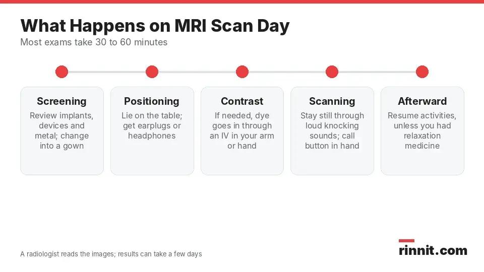
Knowing the sequence of the visit ahead of time can make the experience feel more predictable.
What if I have claustrophobia?
If tight spaces make you anxious, your provider may offer medicine to help you relax during the scan [1]. An open MRI may also be an option [1].
For children, non-drug approaches such as preparation, information and distraction have been linked to less need for sedation or general anesthesia [4].
Does an MRI hurt?
An MRI itself is painless. Some parts of the exam can still be uncomfortable [1]:
Lying still for 30 minutes or longer
The loud noise of the scanner
Feeling closed in, if you are sensitive to tight spaces
A brief pinch if you need an IV for contrast
You may notice mild warmth in the area being scanned. This is normal, but tell the technologist if it bothers you [1].
How long does an MRI take?
Most MRI exams take 30 to 60 minutes. The exact time depends on the body area and the type of images needed, so ask your provider for a more specific estimate [1].
What happens after an MRI, and what do the results mean?
Can I go back to normal activities?
If you did not receive relaxation medicine, you need no recovery time and can return to your usual activities right away [1].
If you did receive relaxation medicine, you will need time to recover and someone to drive you home. Any mild side effects from contrast usually fade quickly [1].
How are MRI results reported?
A radiologist, a doctor trained to read imaging tests, reviews your images for growths, inflammation, injury or other signs of disease. This can take up to a few days. The radiologist then sends a report to your provider, who will explain the findings and any next steps [1].
Result | What it means |
Normal | Nothing concerning was seen in the area that was scanned |
Abnormal | Something unusual was found; this is not always serious, but you may need more tests |
Sources: [1]
There is no single "normal range" for an MRI. What counts as normal depends on the body part and the reason for the scan [1].
What are the risks of an MRI?
MRI is generally low risk when safety guidelines are followed, and its benefits usually outweigh its risks [1]. Possible concerns include:
In one large study of adult inpatients, contrast MRI was not linked to more acute kidney injury, including in people with moderately reduced kidney function [3].
When should I contact my provider?
Call your provider before your scan if you have an implant or device you are unsure about, are or might be pregnant, or are worried about lying still or tight spaces [1]. After your scan, contact your provider if you have questions about your results or the follow-up plan [1].
Conclusion
An MRI gives your care team a detailed, radiation-free view of soft tissues, organs, bones and blood vessels. It helps diagnose and monitor a wide range of conditions, from stroke and multiple sclerosis to joint injuries and heart disease [1].
Your most important job is honest safety screening: report every implant, device and possible metal fragment, remove metal items, and mention pregnancy or claustrophobia so the team can plan a safe, comfortable scan [1].
Keep learning: If small spaces worry you, read What Is Claustrophobia (Fear of Enclosed Spaces)? Symptoms, Causes, Diagnosis & Treatment. If you have a hearing implant, see Cochlear Implants: How They Work, Who They Help, and What Recovery Looks Like.
FAQ
What is an MRI scan?
An MRI is an imaging test that uses a strong magnet, radio waves and a computer to make detailed pictures of soft tissues and organs. It does not use radiation [1].
Why would a doctor order an MRI?
To diagnose or monitor problems such as stroke, multiple sclerosis, tumors, aneurysms, heart disease and joint injuries [1].
How do I prepare for an MRI?
Tell your care team about any implants, devices or metal fragments in your body, and whether you are pregnant or claustrophobic. On the day, remove jewelry, watches, hearing aids, phones and other metal items [1].
Can I have an MRI if I have a pacemaker or other implant?
You must tell your provider and technologist about any pacemaker, defibrillator, stimulator, drug pump, shunt, aneurysm clip, stent or similar implant before the scan. The team will decide whether and how the scan can be done safely [1].
Does an MRI hurt?
No, the scan itself is painless. Lying still, the loud noise and the enclosed space can be uncomfortable, and an IV for contrast may cause a brief pinch [1].
How long does an MRI take?
Most MRI scans take 30 to 60 minutes. Your provider can give you a more exact estimate for your exam [1].
What is MRI contrast, and is it safe?
Contrast is a dye injected through an IV to make tumors, inflammation and blood vessels easier to see. It is generally safe, serious reactions are rare, and mild effects such as nausea or a metallic taste are uncommon [1, 2].
Is MRI safe during pregnancy?
Tell your provider if you are or might be pregnant before any MRI, and especially before contrast. Your provider will weigh whether the scan and contrast are needed [1].
How soon will I get my MRI results?
A radiologist reviews your images and sends a report to your provider, which can take up to a few days. Your provider will then explain the results [1].
What does an abnormal MRI result mean?
An abnormal result means the scan showed something that needs attention. It is not always serious, but your provider may recommend more tests [1].
Related Reading
Stroke: Symptoms, Causes, Diagnosis, and Treatment — A Complete Guide explains stroke warning signs, diagnosis and treatment.
What Is Multiple Sclerosis? A Plain-Language Guide to Symptoms, Types, and How It's Treated covers the symptoms and types of a condition that brain and spine MRI helps diagnose and monitor.
Brain Aneurysm: Symptoms, Causes, and Treatment explains how bulging brain arteries form and how they are treated.
Brain Tumor: Symptoms, Causes, Diagnosis and Treatment — What You Need to Know covers brain tumor signs, diagnosis and treatment.
Herniated Disk: Symptoms, Causes & What Actually Helps explains how a slipped disk presses on nerves and what helps.
O'Brien Test: How It's Done, Positive vs Negative Results and Next Steps explains the quick shoulder exam used to screen for a SLAP tear or an AC joint problem.
Gadolinium Contrast for MRI: How It Works, Side Effects and Kidney Safety explains the MRI contrast dye, its side effects and kidney safety.
References
Cleveland Clinic. MRI (Magnetic Resonance Imaging). Cleveland Clinic. Last updated August 5, 2026. https://my.clevelandclinic.org/health/diagnostics/4876-magnetic-resonance-imaging-mri
Yu OJ, Kim PH, Yoon HM, et al. Safety of Gadolinium-based Contrast Agents in Children: A Systematic Review and Meta-Analysis. Radiology. 2025;316(1):e241224. PMID 40728401. https://doi.org/10.1148/radiol.241224
Han MW, Kim PH, Suh CH, et al. Risk of Acute Kidney Injury Following Gadolinium-based Contrast Agent-enhanced MRI: Propensity-matched 7-year Cohort Study. Invest Radiol. 2026;61(5):334-340. PMID 40977576. https://doi.org/10.1097/RLI.0000000000001239
Thestrup J, Hybschmann J, Madsen TW, et al. Nonpharmacological Interventions to Reduce Sedation and General Anesthesia in Pediatric MRI: A Meta-analysis. Hosp Pediatr. 2023;13(10):e301-e313. PMID 37727937. https://doi.org/10.1542/hpeds.2023-007289
Medical disclaimer
This article is for general education only and does not replace advice from a qualified healthcare professional. Talk to your doctor about your own diagnosis and treatment, and seek emergency care right away for severe or sudden symptoms.



Comments