Understanding Restrictive Cardiomyopathy: Causes, Symptoms, and Heart Health

Updated: 2 days ago
Medically reviewed by Dr. Baraa Alnahhal, MD · Last reviewed: October 2026
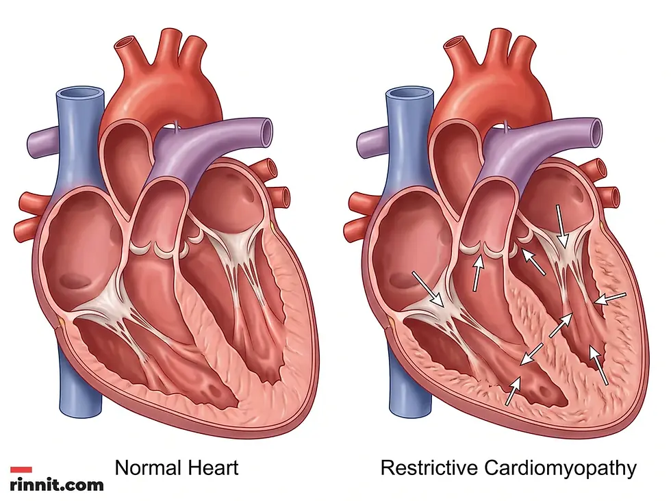
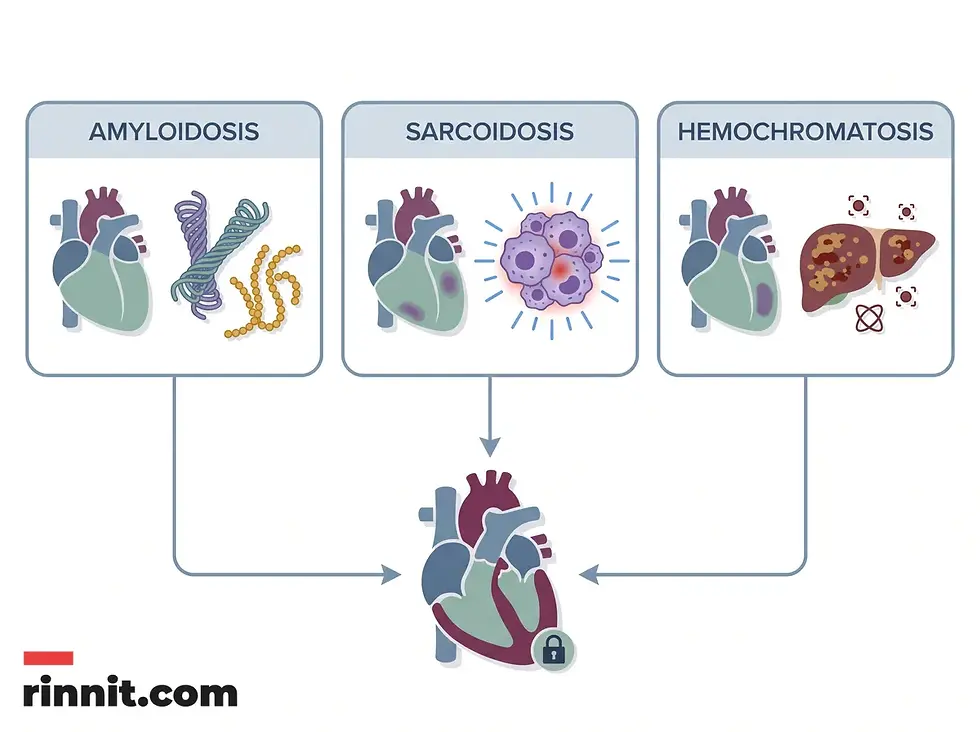
Restrictive cardiomyopathy (RCM) is a rare heart condition in which the lower chambers (ventricles) become stiff and cannot fill properly with blood. [1] This stiffness reduces blood flow and often leads to heart failure. RCM is frequently caused by underlying conditions such as amyloidosis, sarcoidosis or hemochromatosis. [1] [2] Treatment focuses on managing the underlying cause and easing heart failure symptoms with medication and lifestyle changes. [1]
Quick Answer
Restrictive cardiomyopathy is a heart muscle disease in which the ventricles become stiff. [1] This prevents the chambers from filling with enough blood between heartbeats. [1] As blood flow falls, pressure inside the heart rises, which can cause fluid in the lungs and heart failure symptoms. [1] It is the rarest form of cardiomyopathy, accounting for about 5% of all cases. [1]
What the research says
Three treatable causes account for much of the picture, and late recognition is the main barrier to better survival. A review from Mayo Clinic describes restrictive cardiomyopathy as a heterogeneous group of diseases and focuses on cardiac amyloidosis, cardiac sarcoidosis and cardiac hemochromatosis, each of which has tailored treatment but is often diagnosed late (Muchtar et al., Circulation Research, 2017 [2]).
In children, outcomes are poor and heart failure at diagnosis raises risk. In the Pediatric Cardiomyopathy Registry (1990 to 2008, 3,375 children with cardiomyopathy), restrictive cardiomyopathy made up 4.5% of cases; among children with pure restrictive cardiomyopathy, survival was 82%, 80% and 68% at 1, 2 and 5 years, and transplant-free survival was 48%, 34% and 22%. Heart failure at diagnosis was linked to lower transplant-free survival (hazard ratio 2.20) (Webber et al., Circulation, 2012 [3]).
What is restrictive cardiomyopathy and how does it affect heart function?
Restrictive cardiomyopathy is a complex cardiovascular condition in which the muscle of the heart's lower chambers (the ventricles) becomes stiff and rigid. [1] Unlike other forms of heart muscle disease that involve thickening or enlargement of the heart, restrictive cardiomyopathy mainly affects the heart's ability to relax and fill with blood. This impaired filling, known as diastolic dysfunction, forces the heart to work harder and can eventually lead to heart failure.
Cardiomyopathy as a whole is common, affecting up to 1 in 500 people, but the restrictive type is exceptionally rare. [1] It is also called idiopathic restrictive cardiomyopathy (when the cause is unknown) or infiltrative cardiomyopathy (when abnormal substances build up in the heart tissue). [1] Understanding how it disrupts normal blood flow is important for early detection and management.
Feature | Restrictive Cardiomyopathy (RCM) | Normal Heart Function |
Ventricle State | Rigid and stiff | Flexible and elastic |
Blood Filling | Severely limited | Full and efficient |
Heart Rhythm | Rhythm problems (arrhythmia) can occur | Steady and regular |
Blood Flow | Reduced circulation | Optimal circulation |
Main Risk | Heart failure and fluid buildup | None |
What are the symptoms and warning signs of restrictive cardiomyopathy?
Some people with restrictive cardiomyopathy have no symptoms in the early stages. [1] As the heart's ability to fill and pump declines, symptoms typical of heart failure appear, caused by blood and fluid backing up into the lungs and extremities.
Symptoms can include:
Because these symptoms overlap with many other heart and lung conditions, a professional medical evaluation is needed to confirm the diagnosis.
What causes restrictive cardiomyopathy and who is at risk?
Restrictive cardiomyopathy is often the result of an underlying condition that makes the heart muscle stiff. Examples include amyloidosis (protein deposits), hemochromatosis (iron overload) and sarcoidosis (an inflammatory disease), as well as connective tissue diseases. [1] Sarcoidosis is most prevalent in Black females. [1]
Certain cancer treatments also raise the risk. [1] People who have had chemotherapy or radiation therapy may be more likely to develop restrictive cardiomyopathy. [1] Finding the root cause is the first step in building an effective treatment plan.
How is restrictive cardiomyopathy diagnosed?
Healthcare providers begin with a physical examination, listening to the heartbeat and measuring blood pressure. [1] The key test is an echocardiogram, which uses ultrasound to show how well the ventricles fill and pump. [1]
Additional tests help separate restrictive cardiomyopathy from other conditions and identify an underlying cause. These can include blood and urine tests, a chest X-ray, a heart MRI or cardiac CT scan, a heart PET scan, right heart catheterization, and genetic testing. [1] In some cases a myocardial biopsy, in which a small sample of heart tissue is examined, is needed for a definitive diagnosis. [1]
Diagnostic Test | Purpose in RCM Evaluation |
Echocardiogram | Assesses how well the ventricles fill and function |
Heart MRI | Provides detailed images of the heart muscle |
Chest X-ray | Checks for fluid in the lungs or an enlarged heart |
Blood and urine tests | Look for signs of an underlying systemic disease |
Myocardial biopsy | Confirms conditions such as amyloidosis |
How is restrictive cardiomyopathy managed and treated?
No specific treatment reverses the stiffening of the heart muscle in restrictive cardiomyopathy. Instead, care focuses on treating the underlying cause and controlling heart failure symptoms. [1] For example, corticosteroids may be used for sarcoidosis, therapeutic phlebotomy (blood removal) for hemochromatosis, and specific medications for certain types of amyloidosis. [1]
To manage heart failure symptoms and prevent complications, providers may prescribe diuretics to reduce fluid retention and ease the workload on the heart. Medications such as beta-blockers or calcium channel blockers may be used to treat heart rhythm irregularities. [1] Some people ultimately need a heart transplant, and palliative care may also be recommended. [1]

Conclusion
Restrictive cardiomyopathy is a serious but rare condition that needs careful medical management. Although the stiffness of the heart muscle cannot easily be reversed, identifying and treating the underlying cause and controlling heart failure symptoms can improve quality of life and help prevent complications such as arrhythmia and stroke. Working closely with a cardiology team and following the prescribed plan supports long-term heart health.
Next step: If you have persistent fatigue, unexplained swelling or shortness of breath, see a healthcare provider for a heart evaluation. Early diagnosis and management are key to protecting your heart.
Frequently Asked Questions
What is the main difference between restrictive and other cardiomyopathies?
Is restrictive cardiomyopathy hereditary?
Some underlying causes, such as hemochromatosis and certain forms of amyloidosis, can be inherited, and genetic testing may be part of the evaluation. Whether it applies to you depends on the cause, so ask your cardiology team.
Can lifestyle changes help manage restrictive cardiomyopathy?
Lifestyle measures can support treatment but do not replace it. Providers often recommend a sodium-controlled diet, keeping blood pressure under control and exercising as directed, alongside prescribed medication. [1]
What are the most serious complications of restrictive cardiomyopathy?
The main complications are heart failure, heart rhythm problems (arrhythmia), and blood clots (thromboembolism), which can lead to stroke. [1]
How rare is restrictive cardiomyopathy?
Restrictive cardiomyopathy is the rarest type of cardiomyopathy, accounting for about 5% of all cases. Cardiomyopathy overall is common, affecting up to 1 in 500 people. [1]
What is the role of a heart transplant in restrictive cardiomyopathy?
Some people with restrictive cardiomyopathy ultimately need a heart transplant, particularly when heart failure no longer responds well to medication. Your care team can advise on whether this applies to you.
When should I seek emergency care?
Call your local emergency number if you have chest pain, fainting, or sudden or severe shortness of breath. Contact your healthcare provider if heart failure symptoms develop or worsen. [1]
What is infiltrative cardiomyopathy?
Infiltrative cardiomyopathy is another name used for restrictive cardiomyopathy, particularly when abnormal substances such as protein (amyloid) or iron build up in the heart muscle and make it stiff.
Related Reading
Heart MRI (Cardiac MRI): What It Shows, How to Prepare, What Happens and Results explains the imaging test used to assess the heart muscle and its lining.
References
Cleveland Clinic. Restrictive Cardiomyopathy. Cleveland Clinic. Last updated July 20, 2022. https://my.clevelandclinic.org/health/diseases/17427-restrictive-cardiomyopathy
Muchtar E, Blauwet LA, Gertz MA. Restrictive cardiomyopathy: genetics, pathogenesis, clinical manifestations, diagnosis, and therapy. Circulation Research. 2017;121(7):819-837. PMID 28912185. https://doi.org/10.1161/CIRCRESAHA.117.310982
Webber SA, Lipshultz SE, Sleeper LA, et al. Outcomes of restrictive cardiomyopathy in childhood and the influence of phenotype: a report from the Pediatric Cardiomyopathy Registry. Circulation. 2012;126(10):1237-1244. PMID 22843787. https://doi.org/10.1161/CIRCULATIONAHA.112.104638
Medical disclaimer
The information provided in this article is for educational purposes only and should not be used as a substitute for professional medical advice, diagnosis, or treatment. Always seek the advice of your physician or other qualified health provider with any questions you may have regarding a medical condition.



Comments