Echocardiogram With Strain: How Strain Imaging Spots Early Heart Damage

Updated: 3 days ago
Written and medically reviewed by Dr. Baraa Alnahhal, MD · Last reviewed: November 2026 · How we review
Quick Answer
An echocardiogram with strain (strain imaging) is a heart ultrasound that measures how much the heart muscle shortens and lengthens with each beat. It can pick up subtle heart muscle weakness before the ejection fraction drops. It's often used to monitor the heart during chemotherapy and to follow cardiomyopathy and heart failure.
What the research says
Strain spots chemotherapy heart damage early: a systematic review found that an early drop in global longitudinal strain of about 10% to 15% during cancer treatment was the most useful sign for predicting later cardiotoxicity, defined as a fall in ejection fraction or heart failure (Thavendiranathan P et al., Journal of the American College of Cardiology, 2014) [2]
Strain predicts outcomes better than ejection fraction: a meta-analysis across heart failure, heart attack and valve disease found global longitudinal strain was more strongly linked to death than ejection fraction, with about 1.6 times greater predictive strength per standard deviation change (Kalam K et al., Heart, 2014) [3]
Key takeaways
Strain imaging measures how well the heart muscle stretches and contracts.
It can find early heart muscle problems before ejection fraction falls.
It's commonly used to monitor people on heart-affecting cancer treatment.
The test takes about an hour and is painless.
You can eat, drink and take medicines as usual.
What is an echocardiogram with strain?
A standard echocardiogram uses sound waves to show your heart's size, valves and pumping. Strain imaging adds special software that tracks tiny points in the heart muscle and measures how much each segment changes length during the heartbeat. The most common measure is global longitudinal strain (GLS), which reflects how much the heart shortens from top to bottom. Because the heart muscle often starts to weaken in subtle ways first, strain can reveal problems earlier. Sources: [1, 3]
Why it's done

Strain imaging finds subtle changes early.
Screening and follow-up during cancer treatments that can harm the heart, such as some chemotherapy drugs.
Checking the heart after radiation to the chest.
Evaluating and monitoring cardiomyopathy and heart failure, including before and after a biventricular pacemaker.
Assessing pericardial disease. Sources: [1, 2]
Learn more about cardiotoxicity from cancer treatment. Sources: [1]
Why strain matters
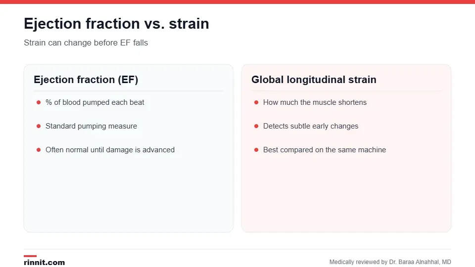
Strain can change before ejection fraction does.
Measure | What it shows | Limitation |
Ejection fraction (EF) | Percentage of blood pumped out with each beat | Often stays normal until damage is advanced |
Global longitudinal strain (GLS) | How much the heart muscle shortens | Values vary somewhat by machine and software |
Because results can differ between machines, providers usually compare your strain over time on the same system. Sources: [1, 2, 3]
What happens during the test
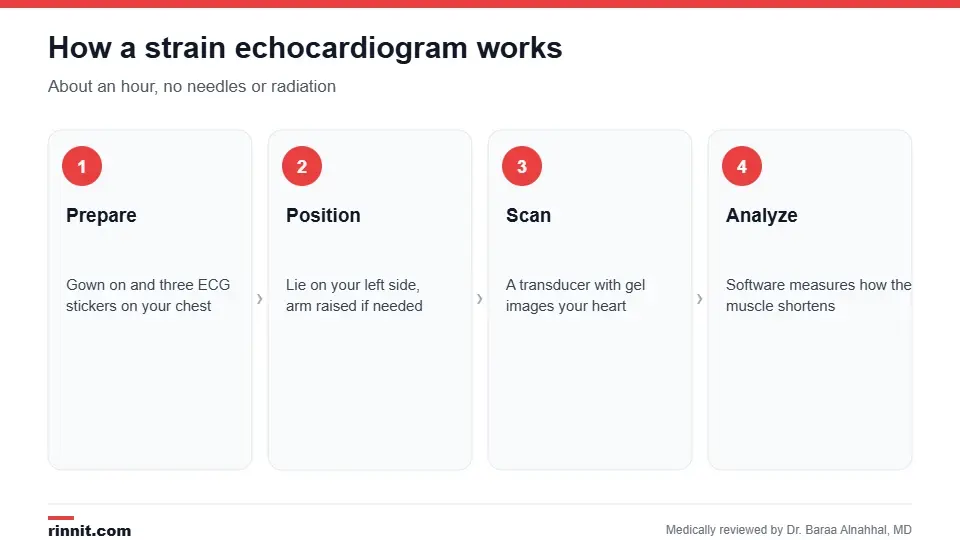
The test takes about an hour.
You change into a gown from the waist up, and three ECG stickers are placed on your chest.
You lie on your left side, sometimes with your left arm raised.
A sonographer moves a transducer with gel over several areas of your chest, asking you to change position or hold your breath at times.
Software analyzes the images to calculate strain. Sources: [1]
You may feel the cool gel and some pressure from the transducer. There are no needles or radiation. You can go home or to other appointments right after. Sources: [1]
Results
A cardiologist reviews the images and adds the results to your medical record. Your provider explains whether your heart muscle function is normal, whether it has changed since earlier tests and whether you need treatment or closer monitoring. During cancer treatment, a meaningful drop in strain may lead your care team to start heart-protecting medicine or adjust your therapy. Sources: [1, 2]
Conclusion
An echocardiogram with strain adds a sensitive measure of heart muscle health to a routine heart ultrasound. It's especially valuable for catching early heart changes during cancer treatment, when acting early can protect your heart.
Keep learning: read about the echocardiogram and cardiac imaging.
Frequently asked questions
What does strain mean on an echocardiogram?
How much your heart muscle shortens and stretches with each beat.
What is global longitudinal strain?
The most common strain measure, showing how much the heart shortens lengthwise.
Why is strain better than ejection fraction?
It can detect heart muscle problems earlier.
Who needs a strain echocardiogram?
Often people on heart-affecting chemotherapy, and those with cardiomyopathy or heart failure.
How long does it take?
About an hour.
Do I need to prepare?
No. Eat, drink and take medicines as usual.
Does it hurt?
No. You may feel cool gel and some pressure.
Is there radiation?
No. It uses sound waves.
Who reads the results?
A cardiologist.
What happens if my strain drops during chemotherapy?
Your care team may start heart-protecting medicine or adjust treatment.
Related Reading
Echocardiogram: Types, What to Expect, Results and Risks the standard heart ultrasound that strain builds on.
Cardiotoxicity: Cancer Treatment and the Heart — Plain-English Guide how cancer treatment can affect the heart.
Heart Failure: Symptoms, Causes & Treatment Guide a condition strain imaging helps monitor.
Cardiac Imaging: Types of Heart Scans and What Each One Shows an overview of all heart imaging tests.
Chemotherapy: How It Works, Side Effects, and What to Expect the treatment that often calls for strain monitoring.
References
Cleveland Clinic. Echocardiogram with Strain Imaging. Cleveland Clinic. Last updated October 31, 2022. https://my.clevelandclinic.org/health/diagnostics/16948-echocardiogram-with-strain-imaging
Thavendiranathan P, Poulin F, Lim KD, et al. Use of myocardial strain imaging by echocardiography for the early detection of cardiotoxicity in patients during and after cancer chemotherapy: a systematic review. Journal of the American College of Cardiology. 2014;63(25 Pt A):2751-2768. PMID 24703918. https://doi.org/10.1016/j.jacc.2014.01.073
Kalam K, Otahal P, Marwick TH. Prognostic implications of global LV dysfunction: a systematic review and meta-analysis of global longitudinal strain and ejection fraction. Heart. 2014;100(21):1673-1680. PMID 24860005. https://doi.org/10.1136/heartjnl-2014-305538
Medical disclaimer
This article is for general education only and does not replace advice from a qualified healthcare professional. Talk to your doctor about your own diagnosis and treatment, and seek emergency care right away for severe or sudden symptoms.



Comments