Eye Exam: What Happens, Tests Included and How Often to Go

Updated: 2 days ago
Written and medically reviewed by Dr. Baraa Alnahhal, MD · Last reviewed: November 2026 · How we review
Quick Answer: What is an eye exam?
An eye exam is a painless, noninvasive check of how well you see and how healthy your eyes are, done by an optometrist or ophthalmologist. A comprehensive exam covers 13 areas, from reading a letter chart to looking at the retina and optic nerve. Most people should have one every one to two years, even with 20/20 vision. [1]
What the research says
More than half of people with glaucoma did not know they had it. Pooled data on 55,415 adults aged 40 and older from 14 European population studies found an age-standardized glaucoma prevalence of 2.99%. Of all cases found by direct eye examination, 56.4% had never been diagnosed, rising to more than 80% in people younger than 55. (Stuart et al., Ophthalmology, 2025) [2]
The leading causes of blindness are mostly conditions an eye exam can pick up. In adults aged 50 and older in 2020, the top causes of blindness worldwide were cataract (15.2 million cases), glaucoma (3.6 million), undercorrected refractive error (2.3 million), age-related macular degeneration (1.8 million) and diabetic retinopathy (0.86 million). Undercorrected refractive error was the leading cause of moderate to severe vision impairment, at 86.1 million cases. (GBD Vision Loss Expert Group, The Lancet Global Health, 2021) [3]
Retinal photos screen well for diabetic eye disease, but in-person exams set the standard. Across 28 studies that compared remote retinal photo screening with a face-to-face eye exam, photos detected any diabetic retinopathy with 91% sensitivity and 88% specificity (13 studies, 7,207 people). Detection of age-related macular degeneration was weaker, at 71% sensitivity. (Mehraban Far et al., BMJ Open Ophthalmology, 2022) [4]
Key takeaways
An eye exam checks both your eyesight and the health of your eyes, and it can find problems before you notice symptoms. [1]
A comprehensive exam has 13 parts, including visual acuity, visual fields, eye pressure, a slit lamp exam of the front and back of the eye, and a check of orientation. [1]
Dilating drops are common. Afterward, close-up focusing is hard for a while and light may feel uncomfortable, so bring sunglasses and consider a driver. [1]
The only listed risk is an allergic reaction to the dilating drops, and side effects are rare and usually minor. [1]
Most people need an exam every one to two years; people over 60, people with diabetes and others at higher risk need them more often. [1]
Silent disease is common: in one large analysis, 56.4% of people with glaucoma had not been diagnosed before. [2]
What is an eye exam?
An eye exam is a series of simple tests that show how well your eyes work and whether there are signs of eye disease. It is noninvasive, which means nothing is inserted into your body. [1]
Think of it as a yearly checkup for your eyes. Everyone needs one, not only people who wear glasses or contact lenses, because many eye problems cause no symptoms early on. [1]
Two types of eye specialists perform eye exams. A third, the optician, works with glasses but does not examine your eyes. [1]
Professional | Training | What they do |
Ophthalmologist | MD or DO degree: 4 years of medical school plus 4 years of residency, sometimes a fellowship | Diagnoses and treats eye conditions; many also perform eye surgery |
Optometrist | OD degree: 4 years of optometry school, sometimes a 1-year residency | Diagnoses and treats eye problems; does not perform surgery |
Optician | Not an eye exam provider | Helps you choose glasses, takes eye and head measurements, fits and adjusts frames |
Sources: [1]
Why is an eye exam done?
The main goal is to protect your sight. An exam checks how clearly you see, measures whether you need corrective lenses, and looks for early signs of eye disease such as glaucoma. [1]
Eye exams fall into three broad groups, based on why you are going: [1]
Routine (general) exams: Regular checkups for people with no known eye condition, or who only need glasses or contacts. [1]
Follow-up exams: Visits after an eye injury or eye procedure, or ongoing checks for a long-term condition such as diabetes or macular degeneration. [1]
Medical or emergency exams: Urgent checks for problems that could threaten your vision, such as an eye injury, sudden vision loss or a head injury. [1]
Research shows why routine checks matter. Worldwide, cataract, glaucoma, undercorrected refractive error (vision that glasses or contacts could improve), macular degeneration and diabetic retinopathy were the leading causes of blindness in adults aged 50 and older in 2020. [3]
What does a comprehensive eye exam check?
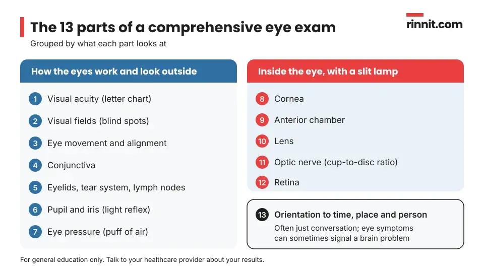
A comprehensive eye exam has 13 parts. Seven look at how your eyes work and how the outside of the eye looks. Five use a slit lamp, a microscope with a bright light, to look inside the eye. The last part checks your orientation. [1]
Part of the exam | What it checks |
Visual acuity | How clearly you see, usually with a Snellen chart of letters or symbols that shrink line by line |
Visual fields | Each section of your side and central vision in each eye, looking for gaps or blind spots |
Movement and alignment | Whether your eyes line up and move in every direction, one eye at a time and then together |
Conjunctiva | The thin layer over the white of the eye and the inside of the eyelids |
Ocular adnexa | Eyelids, tear system and the lymph nodes in front of the ears |
Pupil and iris | How your pupils react to light (pupillary reflexes) |
Intraocular pressure | Fluid pressure inside the eye, often measured with a puff of air; high readings can point to glaucoma |
Cornea (slit lamp) | The condition of the clear front window of the eye |
Anterior chamber (slit lamp) | The fluid-filled space at the front of the eye, iris and lens; helps spot angle-closure glaucoma |
Lens (slit lamp) | Whether the lens is clear and normally shaped |
Optic nerve (slit lamp) | The optic disc and cup, including the cup-to-disc ratio |
Retina (slit lamp) | Wrinkles, color changes or damage to the light-sensing layer at the back of the eye |
Orientation | Whether you know the date, where you are and details about yourself |
Sources: [1]
The orientation check is usually just conversation. It matters because eye symptoms sometimes signal a problem in the brain. [1]
If you need glasses or contacts, your specialist also measures the refractive error (how far your eye's focus is from normal) in each eye to write a prescription. Contact lens wearers have extra measurements of the eyeball. [1]
How do you prepare for an eye exam?
Routine and follow-up exams need little preparation. A few simple steps help your specialist get the full picture: [1]
Bring a complete list of your medicines, vitamins and supplements, including those for conditions that seem unrelated to your eyes. [1]
Bring your current glasses or contact lenses and any previous prescriptions. [1]
Pack sunglasses, because dilated eyes can be sensitive to light. [1]
Arrange a ride if you think you may be dilated, since some people have trouble seeing well enough to drive afterward. [1]
Tell your specialist about any past reaction to eye drops. [1]
What happens during an eye exam?
Your visit usually follows a steady order, though your specialist may add tests based on what they find. [1]
History: Your specialist asks about your vision, any recent changes, and medical events or life changes. [1]
Vision and function tests: You read a letter chart, follow targets with your eyes and have your side vision tested. [1]
Outer eye checks: Your eyelids, tear system, conjunctiva and pupils are examined. [1]
Eye pressure: A quick puff of air measures the pressure inside your eye. [1]
Dilation and slit lamp exam: Drops relax the muscle that controls your pupil so it widens, making it easier to see the lens, optic nerve and retina. [1]
Refraction, if needed: Your specialist measures each eye's focusing error to write a prescription. [1]
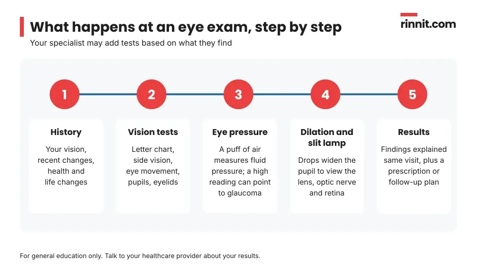
The whole exam is noninvasive, and nothing is placed inside your body. The main after-effect is temporary light sensitivity and blurry near vision if your pupils were dilated. [1]
What happens after an eye exam?
Your specialist explains what they found and what it means for you. If you need corrective lenses, you get a prescription. [1]
In the US, federal rules require your provider to give you a paper or digital copy of your prescription, even if you do not ask. Some states require your pupillary distance (the space between your pupils) on the paper copy, and some providers will share it on request. [1]
If your pupils were dilated, close-up focusing will be difficult for a while, and bright light and screens may hurt. Sunglasses help, and having someone drive you home is a good idea. [1]
What do eye exam results mean?
Unlike many tests, you usually hear your results the same day. Your specialist typically talks through the findings during or right after the exam. [1]
Results may include a new or updated prescription, reassurance that your eyes look healthy, or a recommendation for treatment, more tests or a follow-up visit. For example, a high eye pressure reading can point to glaucoma, and a low cup-to-disc ratio of the optic nerve may point to a higher risk of conditions such as ischemic optic neuropathy (damage to the optic nerve from poor blood flow). [1]
An exam can find disease you had no idea about. In a large European analysis, 56.4% of people found to have glaucoma had never been diagnosed, and the share was above 80% in adults younger than 55. [2]
Exams also matter for people with diabetes. Remote retinal photos caught any diabetic retinopathy with 91% sensitivity compared with a face-to-face eye exam, but were less reliable for macular degeneration, at 71% sensitivity. [4]
Are there any risks or side effects?
Eye exams are very safe. The only listed risk is an allergic reaction to the dilating drops, and side effects are rare and usually minor. [1]
If you have reacted to eye drops before, tell your specialist. They may be able to use an alternative. [1]
How often should you get an eye exam?
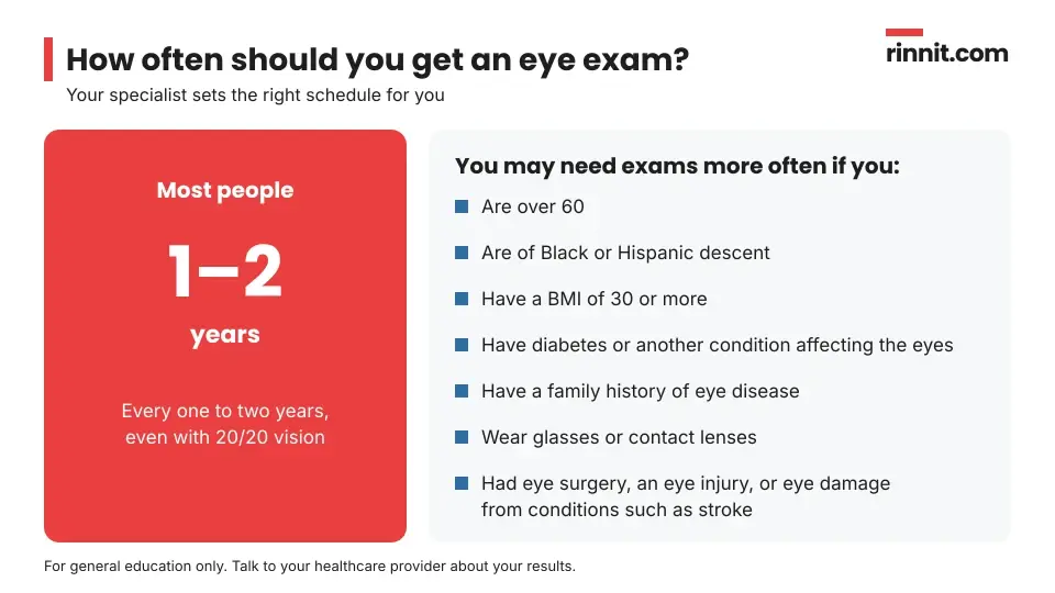
Most people should have a comprehensive eye exam every one to two years, even if they see 20/20. [1]
Your specialist may recommend more frequent exams if you: [1]
Higher-risk group | Examples |
Age | Over 60 |
Background | Black or Hispanic descent |
Body weight | BMI (body mass index) of 30 or more |
Health conditions | Diabetes or another condition that can affect the eyes |
Family history | Relatives with eye disease |
Corrective lenses | Wear glasses or contact lenses |
Eye history | Past eye surgery, eye injury, or eye damage from conditions such as stroke |
Sources: [1]
When should you call your eye specialist?
Book a visit if you notice gradual changes in your vision, such as: [1]
Sudden vision loss is a medical emergency. Get emergency care right away for partial or total vision loss in one or both eyes, including blurring or fading, darkening or dimming, a dark curtain over part or all of your vision, or bright flashes. [1]
Some conditions, such as migraines, can cause short-lived vision loss. If you have one of these, ask your provider ahead of time how to respond when it happens. [1]
Questions to ask your provider
How often should I have an eye exam, given my age and health?
Will my pupils be dilated today, and how long should I avoid driving?
Was my eye pressure normal, and am I at risk for glaucoma?
Did you see any changes in my optic nerve or retina?
Has my prescription changed, and can I have a copy with my pupillary distance?
Do I need any extra tests or a follow-up visit?
What symptoms should make me call you before my next exam?
Conclusion
An eye exam is one of the simplest ways to protect your sight. In a single visit, your specialist checks how clearly you see, measures your eye pressure and looks inside your eye for early signs of disease, often before any symptoms appear. [1]
Plan for an exam every one to two years, or more often if you have diabetes, are over 60 or have other risk factors. Bring your medicine list and glasses, pack sunglasses in case of dilation, and never wait out sudden vision loss. [1]
Keep learning: Read our guides to glaucoma, cataracts and diabetic retinopathy. For more, see the National Eye Institute's page on getting a dilated eye exam.
FAQ
What is an eye exam?
An eye exam is a noninvasive set of tests that checks how well you see and how healthy your eyes are. It is done by an optometrist or ophthalmologist. [1]
Why do I need an eye exam if I see well?
Many eye problems cause no symptoms at first. Routine exams can find them early, even if you have 20/20 vision. [1]
How do I prepare for an eye exam?
Bring a list of your medicines and supplements, your glasses or contacts and past prescriptions. Pack sunglasses and consider a driver in case you are dilated. [1]
Does an eye exam hurt?
No. The exam is noninvasive. If your pupils are dilated, light and screens may feel uncomfortable and close-up focusing may be hard for a while afterward. [1]
How long does an eye exam take?
It depends on how many tests you need and whether your pupils are dilated. Plan extra time afterward, because close-up focusing stays difficult for a while after dilation. [1]
What does a comprehensive eye exam include?
It has 13 parts, including visual acuity, visual fields, eye movement, pupils, eye pressure, a slit lamp exam of the cornea, lens, optic nerve and retina, and an orientation check. [1]
When will I get my eye exam results?
Usually right away. Your specialist typically explains the findings during or just after the exam, along with any prescription or follow-up plan. [1]
Are there risks from eye dilation?
The only listed risk is an allergic reaction to the dilating drops. Side effects are rare and usually minor. [1]
How often should I get an eye exam?
Most people need one every one to two years. You may need exams more often if you are over 60, have diabetes, wear glasses or have a family history of eye disease. [1]
When should I see an eye doctor urgently?
Sudden partial or total vision loss, a dark curtain over your vision or bright flashes are a medical emergency. Seek care right away. [1]
Related Reading
Progressive Lenses: Types, How They Work and Adjusting explains progressive lenses for presbyopia.
Cataract Surgery: What to Expect, Recovery and Results explains how cataract surgery restores clear vision.
Corneal Pachymetry: Measuring Corneal Thickness for Glaucoma and LASIK explains the test that measures corneal thickness.
PERRLA Eye Exam: What It Means and What Pupil Changes Can Show explains the quick pupil check used in eye and neurological exams.
Hirschberg Test (Corneal Light Reflex): How Eye Alignment Is Checked explains the light test used to check eye alignment.
Amsler Grid Eye Test: How to Check Your Central Vision at Home explains the at-home grid used to check central vision.
Learn more in our guide to Glaucoma: The Silent Threat to Vision, and How Early Treatment Can Protect Your Sight.
Learn more in our guide to Cataracts: Symptoms, Causes, Diagnosis and Treatment — What You Need to Know.
Learn more in our guide to Diabetic Retinopathy Explained: How Diabetes Damages the Eyes and What Stops the Damage.
Learn more in our guide to Astigmatism: Symptoms, Causes & Treatment Options — Complete Guide.
Learn more in our guide to Presbyopia: Complete Guide to Age-Related Near Vision Loss, Symptoms, and Treatment Options.
Learn more in our guide to Farsightedness (Hyperopia): Why Close Objects Look Blurry — Causes, Eye Exams, and Your Correction Options.
Fundoscopy (Ophthalmoscopy): What It Is, Types, What to Expect and What It Can Find explains the exam of the retina and optic nerve used in eye and diabetes care.
Gonioscopy: What It Is, What to Expect and What Open or Closed Angles Mean explains the lens exam that shows whether the eye's drainage angle is open or closed.
Visual Acuity Test (Snellen Chart): How It Works and What 20/20 Means explains the eye-chart test that measures how sharply you see.
Slit Lamp Exam: What It Checks, How It's Done and What to Expect explains the microscope eye exam used to check every part of the eye.
Depth Perception Test (Stereopsis Test): Types, How It's Done and What Results Mean explains the test that checks how well the eyes work together to judge depth.
Retinal Imaging: Fundus Photos, OCT and Angiography Explained explains the photos and scans used to check the back of the eye.
Retinoscopy: How Eye Doctors Measure Your Prescription explains the light-reflex eye test used to measure a glasses prescription.
Corneal Topography: Mapping the Cornea and Reading the Results explains the eye test that maps the shape of the cornea.
Keratometry: Measuring Corneal Curve for Astigmatism and Cataract Surgery explains the test that measures the curve of the cornea.
References
Cleveland Clinic. Eye Exam (Eye Test): Vision Test & Other Optical Exams. Cleveland Clinic. Last updated August 5, 2024. https://my.clevelandclinic.org/health/diagnostics/eye-exam
Stuart KV, de Vries VA, Schuster AK, et al. Prevalence of Glaucoma in Europe and Projections to 2050: Findings from the European Eye Epidemiology Consortium. Ophthalmology. 2025;132(10):1114-1124. PMID 40499787. https://doi.org/10.1016/j.ophtha.2025.06.002
GBD 2019 Blindness and Vision Impairment Collaborators; Vision Loss Expert Group of the Global Burden of Disease Study. Causes of blindness and vision impairment in 2020 and trends over 30 years, and prevalence of avoidable blindness in relation to VISION 2020: the Right to Sight: an analysis for the Global Burden of Disease Study. Lancet Glob Health. 2021;9(2):e144-e160. PMID 33275949. https://doi.org/10.1016/S2214-109X(20)30489-7
Mehraban Far P, Tai F, Ogunbameru A, et al. Diagnostic accuracy of teleretinal screening for detection of diabetic retinopathy and age-related macular degeneration: a systematic review and meta-analysis. BMJ Open Ophthalmol. 2022;7(1):e000915. PMID 35237724. https://doi.org/10.1136/bmjophth-2021-000915
Medical disclaimer
This article is for general education only and does not replace advice from a qualified healthcare professional. Talk to your doctor about your own diagnosis and treatment, and seek emergency care right away for severe or sudden symptoms.



Comments