Fundoscopy (Ophthalmoscopy): What It Is, Types, What to Expect and What It Can Find

Updated: 2 days ago
Written and medically reviewed by Dr. Baraa Alnahhal, MD · Last reviewed: November 2026 · How we review
Quick Answer
Fundoscopy (ophthalmoscopy) is an exam of the back of the eye: the retina, its blood vessels, the macula and the optic nerve. A provider shines a light through the pupil with a scope, sometimes after dilating drops. It takes a few minutes, is painless, and can reveal diabetic retinopathy, glaucoma damage, retinal detachment and macular degeneration.
What the research says
Retinal photography works with or without dilating drops: a meta-analysis of 80 studies and more than 50,000 participants found fundus photography detected diabetic retinopathy with 90% sensitivity after dilation versus 89% without (specificity 90% vs 85%), and sensitivity climbed from 0.82 to 0.98 as more retinal fields were photographed (Dattilo V et al., European Journal of Ophthalmology, 2026) [2]
Handheld cameras extend fundoscopy to the emergency room: a best-evidence review of 12 papers concluded that portable handheld fundus cameras are effective for diagnosing diabetic retinopathy in emergency settings; in the strongest study, a handheld camera with AI reading reached 82.1% sensitivity and 97.4% specificity (Arzaghi M, Alavi-Moghaddam M, Emergency Medicine Journal, 2024) [3]
Key takeaways
Looks directly at the retina, optic disc, macula and retinal vessels.
Direct, indirect, monocular indirect and slit-lamp techniques give different views; fundus photography records the image.
Dilating drops are optional but widen the view; retinal photography performs almost as well without them.
No risks; mild glare and after-images are normal.
Part of routine eye exams, diabetes care, blood pressure checks and headache work-ups.
What is fundoscopy?
The fundus is the inner back surface of the eye. It is the only place in the body where a provider can see blood vessels and a nerve (the optic nerve) directly, without surgery or imaging. That is why fundoscopy is used not only by eye specialists but also by primary care, emergency and neurology clinicians. Sources: [1]

Fundoscopy takes only a few minutes and is painless.
Types of ophthalmoscopy
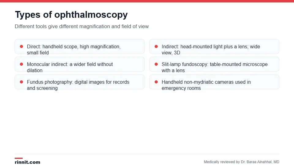
Different scopes trade magnification for field of view.
Type | How it is done | Best for |
Direct | Handheld scope held close to your eye; ~15x magnification, small field | Quick checks of the optic disc and macula |
Indirect | Head-mounted light plus a handheld lens; wide, 3D view | Peripheral retina, retinal tears and detachment |
Monocular indirect | Handheld device with a wider field; works without dilation | Screening in clinics and emergency rooms |
Slit-lamp fundoscopy | Table-mounted microscope with a lens | Detailed, magnified assessment by an eye specialist |
Fundus photography | Digital camera records the retina | Diabetic retinopathy screening, tracking change over time |
Sources: [1]
Why is it done?
Routine eye and physical exams.
Screening and monitoring for diabetic retinopathy.
Assessing the optic nerve in glaucoma.
Checking for hypertensive retinopathy in people with high blood pressure.
Sudden vision loss, flashes or floaters that could signal retinal detachment.
Headache with suspected raised pressure in the brain (papilledema).
Macular degeneration, retinal vein occlusion, infections and some genetic eye diseases. Sources: [1]
How to prepare
No special preparation is needed. If dilating drops are planned, bring sunglasses and arrange a ride, because your near vision will be blurry and your eyes light-sensitive for 4–6 hours. Tell your provider if you have had angle-closure glaucoma or an allergy to eye drops. Sources: [1]
What happens during the exam?
Your provider may put in dilating drops, which take 15–20 minutes to work.
The room is dimmed and you are asked to look at a fixed point.
The provider shines the scope's light into your eye and views the fundus, checking the red reflex, optic disc, vessels, macula and periphery.
Each eye is examined; you may be asked to look in different directions.
If something needs a closer look, fundus photographs, optical coherence tomography (OCT) or a slit-lamp exam may follow.
The bright light can be uncomfortable for a moment, but the exam does not hurt and there are no risks. Sources: [1]
Understanding your results
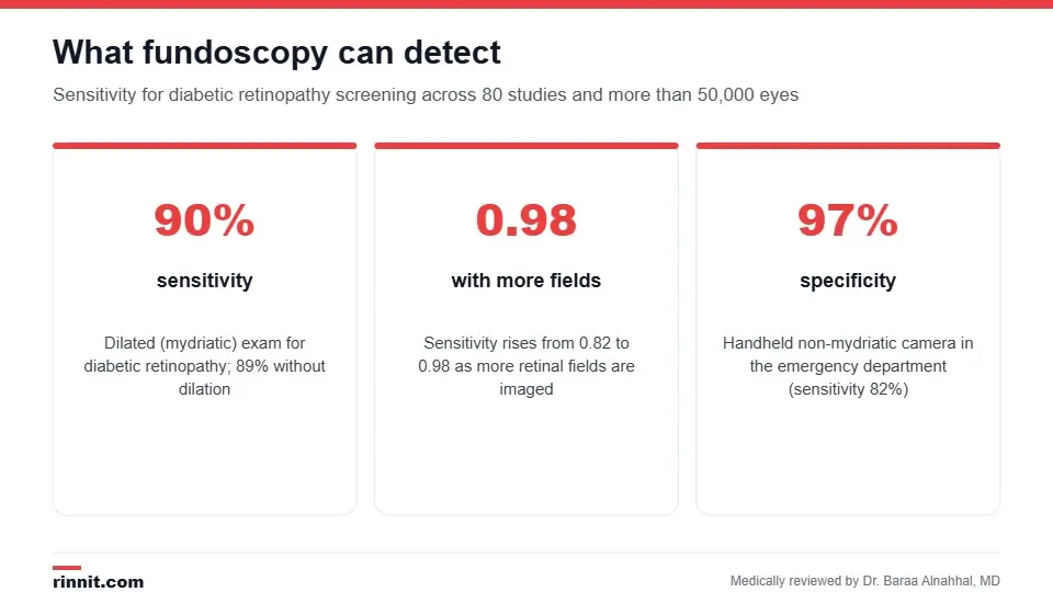
Pooled accuracy for diabetic retinopathy screening from 80 studies.
Finding | What it may indicate |
Microaneurysms, hemorrhages, exudates | Diabetic retinopathy |
Narrowed arteries, nicking where vessels cross | Hypertensive retinopathy |
Large cup-to-disc ratio | Glaucoma |
Swollen, blurred optic disc | Papilledema (raised intracranial pressure) or optic neuritis |
Drusen, pigment changes at the macula | Macular degeneration |
Retinal tear or lifted retina | Retinal detachment |
Research supports fundus imaging as a reliable screening tool. Across 80 studies, retinal photography detected diabetic retinopathy with 90% sensitivity after dilation and 89% without; photographing more retinal fields pushed sensitivity to 0.98. In emergency departments, handheld non-mydriatic cameras reached 82% sensitivity and 97% specificity for diabetic retinopathy. Sources: [2, 3]
Risks
There are none from the exam itself. Dilating drops can cause temporary blurred vision, glare and, very rarely, a rise in eye pressure in people with narrow drainage angles. Sources: [1]
Conclusion
Fundoscopy gives your provider a direct window onto your retina and optic nerve in a few painless minutes. For people with diabetes or high blood pressure it is one of the most valuable routine checks available, and in an emergency it can reveal pressure on the brain or a detaching retina. Ask how often you should have the exam and whether dilation is recommended for you.
Keep learning: see what a full eye exam includes and how glaucoma tests assess the optic nerve.
Frequently asked questions
What is the difference between fundoscopy and ophthalmoscopy?
They are the same exam. "Fundoscopy" refers to looking at the fundus; "ophthalmoscopy" refers to the instrument, the ophthalmoscope.
Do I need dilating drops?
Not always. Drops give a wider view, especially of the peripheral retina, but research shows undilated retinal photography for diabetic retinopathy is almost as sensitive.
Does fundoscopy hurt?
No. The light is bright for a moment, and dilating drops may sting briefly, but the exam is painless.
How long does it take?
The exam itself takes a few minutes per eye. Add 15–20 minutes if dilating drops are used.
Can I drive after a dilated exam?
It is safer not to. Your near vision will be blurry and your eyes light-sensitive for several hours.
What can fundoscopy detect?
Diabetic and hypertensive retinopathy, glaucoma damage, macular degeneration, retinal detachment, optic nerve swelling and some infections and inherited retinal diseases.
How accurate is it for diabetic retinopathy?
Pooled sensitivity is about 90% with dilation and 89% without, with specificity of 85–90%.
What is fundus photography?
A digital photograph of the retina. It records the exam for comparison over time and is widely used in diabetic eye screening programs.
How often should I have the exam?
People with diabetes are usually advised to have a dilated retinal exam at least once a year; others follow the schedule set by their eye care provider.
What happens if something abnormal is found?
You may have fundus photographs, OCT scanning or a slit-lamp exam, and be referred to an ophthalmologist for treatment.
Related Reading
Eye Exam: What Happens, Tests Included and How Often to Go the full examination that fundoscopy is part of.
Diabetic Retinopathy: Symptoms, Causes, Diagnosis & Treatment — Complete Guide the leading condition screened for with fundoscopy.
Glaucoma Tests: Types, What to Expect and What Results Mean how the optic nerve is assessed in glaucoma.
Retinal Detachment: Symptoms, Causes, Diagnosis & Treatment — Complete Guide the emergency that indirect ophthalmoscopy helps identify.
Papilledema: Causes, Symptoms, and Emergency Care optic nerve swelling that fundoscopy detects.
References
Cleveland Clinic. Fundoscopy (Ophthalmoscopy): What it Is & Who Needs It. Cleveland Clinic. Last updated March 12, 2025. https://my.clevelandclinic.org/health/diagnostics/fundoscopy-ophthalmoscopy
Dattilo V, Polito MS, Parravano M, et al. Comparative diagnostic accuracy of photographic methods for detecting diabetic retinopathy: a systematic review and meta-analysis. European Journal of Ophthalmology. 2026;36(2):448-460. PMID 41362080. https://doi.org/10.1177/11206721251406434
Arzaghi M, Alavi-Moghaddam M. Best evidence topic report: are portable handheld fundus cameras effective in diagnosing diabetic retinopathy in emergency settings? Emergency Medicine Journal. 2024;41(12):764-766. PMID 39147563. https://doi.org/10.1136/emermed-2024-214236
Medical disclaimer
This article is for general education only and does not replace advice from a qualified healthcare professional. Talk to your doctor about your own diagnosis and treatment, and seek emergency care right away for severe or sudden symptoms.



Comments