Glaucoma Tests: Types, What to Expect and What Results Mean

Updated: 2 days ago
Written and medically reviewed by Dr. Baraa Alnahhal, MD · Last reviewed: November 2026 · How we review
Quick Answer: What are glaucoma tests?
Glaucoma tests are a set of quick, painless eye exams that look for glaucoma, a group of eye disorders that damage the optic nerve. They measure eye pressure, check the drainage angle and corneal thickness, examine the optic nerve and test side vision. Most results are ready during the same visit. [1]
What the research says
Optic nerve imaging helps, but it is not perfect on its own. A review of 106 studies covering 16,260 eyes found that OCT measurement of the nerve fiber layer picked up about 72% of glaucoma cases while correctly clearing about 93% of people without it. In a group of 200 people with glaucoma, the best imaging measures would still miss about 60, which is why doctors combine several tests. (Michelessi et al., Cochrane Database of Systematic Reviews, 2015) [2]
Corneal thickness matters for people with high eye pressure. An expert panel that reviewed 37 studies found strong and consistent evidence that a thinner central cornea raises the risk of moving from high eye pressure (ocular hypertension) to primary open-angle glaucoma. It advised measuring corneal thickness in everyone with ocular hypertension, since it also affects pressure readings. (Dueker et al., Ophthalmology, 2007) [3]
Computer analysis of eye scans may help find closed drainage angles. Across 6 studies with 5,269 patients, deep learning programs reading anterior segment OCT images detected angle closure with 94% sensitivity and 93.6% specificity compared with gonioscopy. The authors suggest this could help screening where experienced specialists are scarce. (Olyntho et al., Journal of Glaucoma, 2024) [4]
Key takeaways
Glaucoma damages the optic nerve, is usually linked to raised eye pressure and can lead to vision loss or blindness if it is not found and treated. [1]
Testing may involve one test or a combination: an angle exam, corneal thickness test, dilated eye exam, eye pressure check, optic nerve imaging and a visual field test. [1]
The tests are quick, painless and noninvasive, and most results are available at the same appointment. [1]
Eye pressure greater than 22 mmHg is one of the most important signs of glaucoma, but your doctor weighs all results together. [1]
Glaucoma may cause no symptoms until later stages, so regular eye exams are key if you are at risk. [1]
What are glaucoma tests?
Glaucoma is a group of eye disorders that damage the optic nerve, the nerve that carries visual signals from the retina to the brain. It is usually linked to raised pressure inside the eye, tends to get worse over time and can cause vision loss or blindness. [1]
Glaucoma tests are the exams an eye doctor uses to look for this damage and to judge your risk. You may have a single test or several. An ophthalmologist, a physician who specializes in preventing, diagnosing and treating eye disorders, performs and reviews them. [1]
Why are glaucoma tests done?
Your provider may order glaucoma tests if they suspect glaucoma or if you have symptoms such as: [1]

Blind spots or blurry vision
Eye pain or pressure
Headaches
Seeing rainbow-colored rings (halos) around lights
Red eyes
Tunnel vision (loss of side vision)
Testing is also important for people with risk factors, because glaucoma can cause no symptoms until its later stages. [1]
Who is at higher risk of glaucoma?
Risk factor | Why it matters |
Family history of glaucoma | Significantly raises your lifetime risk |
Being Black | Linked to open-angle glaucoma |
East Asian ancestry | Linked to closed-angle glaucoma |
Farsightedness (hyperopia) | Linked to closed-angle glaucoma |
Past eye injury or complicated eye surgery | Raises your risk |
Diabetes or high blood pressure | Raises your risk |
Past or current corticosteroid use, such as prednisone | Raises your risk |
Age over 40 | Risk rises with age |
Sources: [1]
How often should you be tested?
For people at risk of glaucoma, the American Academy of Ophthalmology recommends regular testing based on age: [1]
Age | Recommended testing for people at risk |
40 to 54 | Every 1 to 3 years |
55 to 64 | Every 1 to 2 years |
65 and older | Every 6 to 12 months |
Sources: [1]
What are the types of glaucoma tests?
There are six main glaucoma tests. Each one looks at a different part of the eye, and your ophthalmologist may use one or a combination. [1]
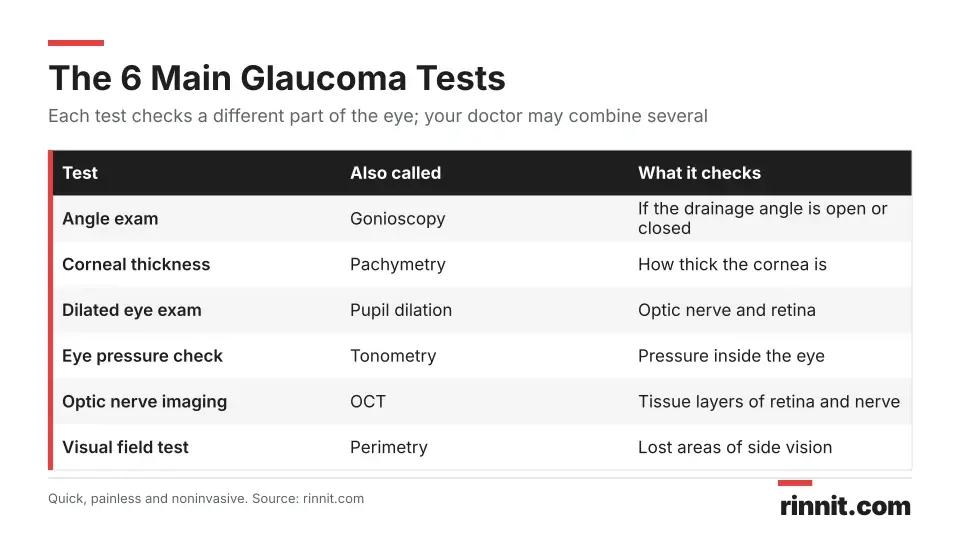
Test | Also called | What it checks | What happens |
Angle exam | Gonioscopy | Whether the drainage angle, where the cornea meets the iris, is open or closed | After numbing drops, a special lens touches the front of your eye |
Corneal thickness measurement | Pachymetry | How thick your cornea is, to help estimate glaucoma risk | After numbing drops, a small probe touches your eye |
Dilated eye exam | Pupil dilation | The color, size, shape and blood vessels of the optic nerve and retina | Drops widen your pupils and a bright, magnifying instrument looks at the back of the eye |
Eye pressure check | Tonometry or applanation | The pressure inside your eye | After numbing drops, a small instrument gently flattens the cornea |
Optic nerve imaging | Optical coherence tomography (OCT) | Each tissue layer of the retina and optic nerve | You rest your chin on a machine and look into a lens; nothing touches the eye |
Visual field test | Perimetry | Lost vision in specific areas, especially side vision | You report what you see while looking straight ahead, or track lights in a machine |
Sources: [1]
Angle exam (gonioscopy)
This test shows whether the angle where your cornea meets your iris is open or closed. A closed angle means the eye's drainage system is blocked, which may point to glaucoma. [1]
Corneal thickness measurement (pachymetry)
A small probe measures how thick your cornea is. A thin cornea raises the risk of primary open-angle glaucoma, so this result helps your doctor estimate your risk. [1] [3]
Dilated eye exam
Drops widen (dilate) your pupils so your doctor can see the back of your eye. A special instrument with a bright light magnifies the retina and optic nerve, letting your doctor judge whether the nerve is damaged and whether glaucoma is the cause. [1]
Eye pressure check (tonometry)
Raised eye pressure is one of the most important signs of glaucoma. The test does not hurt and takes only a few minutes, but you need to stay relaxed and still. [1]
Optic nerve imaging (OCT)
After your pupils are dilated, a digital camera takes pictures of the retina and optic nerve. Many doctors use OCT, which shows each tissue layer and maps the area. Scanning takes about one to two minutes. [1] [2]
Visual field test (perimetry)
This test checks for areas where vision is lost. You may cover one eye at a time and report your side vision while looking straight ahead, look into a machine and respond to lights (automated static perimetry), or view a line pattern and report any blurry or blank areas. Repeating the test over time shows how severe glaucoma is and how fast it is progressing. [1]
How do you prepare for glaucoma tests?
Glaucoma tests do not usually need special preparation. Because some tests use drops that dilate your pupils and can blur your vision, it helps to plan ahead: [1]
Arrange a ride. Your vision is usually blurry afterward, so someone else should drive you home.
Bring sunglasses. Dilated pupils are sensitive to light and need protection from bright light and UV rays.
Share your history. Tell your provider about symptoms and risk factors such as family history, diabetes, eye injuries or steroid use.
Sources: [1]
What happens during glaucoma tests?
The tests are done in your eye doctor's office. The exact tests and their order depend on what your doctor needs to check, but a visit often follows these steps: [1]
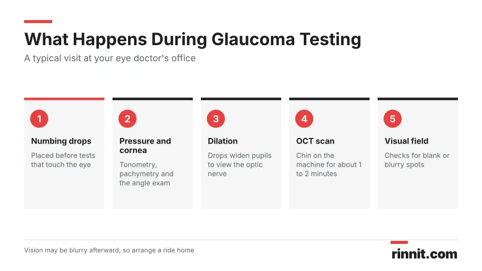
Numbing drops are placed in your eyes before tests in which an instrument touches the eye, such as the eye pressure check, pachymetry and the angle exam.
A small instrument gently flattens your cornea to measure eye pressure while you stay relaxed and still.
A probe measures corneal thickness, and a special lens is placed on the eye to view the drainage angle.
Drops dilate your pupils so the doctor can examine the optic nerve and retina with a bright, magnifying light.
For OCT imaging, you rest your chin on the machine and look into a lens for a scan of about one to two minutes.
A visual field test checks for blank or blurry spots in your vision.
Your ophthalmologist reviews the findings, usually during the same appointment.
Sources: [1]
What happens after glaucoma tests?
After the drops, your vision is usually blurry and your eyes may be sensitive to light. This improves through the day. Wear sunglasses after dilation, have someone drive you home and follow any instructions your doctor gives you. [1]
What do glaucoma test results mean?
Your ophthalmologist considers all your test findings together to decide whether you are at risk, whether you have glaucoma and what type it is. Findings that may point to glaucoma or the need for more testing include: [1]
Test | Finding that may suggest glaucoma |
Angle exam | A narrow or blocked drainage angle |
Corneal thickness | A thin cornea, which raises the risk of primary open-angle glaucoma |
Dilated eye exam | Blood vessels inside the eye with an unusual size or shape |
Eye pressure check | Intraocular pressure greater than 22 mmHg |
Optic nerve imaging | Swelling or buckling of the optic nerve, or a bumpy retina |
Visual field test | Specific spots where vision goes blank or blurry |
Sources: [1]
Cutoffs and reference values can vary between clinics and devices, so ask your doctor what your own numbers mean.
What happens if you have glaucoma?
Glaucoma can be treated and its progression slowed, especially when it is caught early. Depending on your results, next steps may include regular follow-up visits and: [1]
Medicines that lower eye pressure, as eye drops or pills
Laser treatment to improve fluid drainage
Surgery to create a new drainage opening
Surgery to implant drainage tubes
Sources: [1]
What are the risks of glaucoma tests?
Glaucoma tests are safe and carry no known risks. Afterward, your vision may be blurry or sensitive to light, but this improves through the day. [1]
When should you call your provider?
Contact your eye care provider and ask about glaucoma testing if you notice: [1]
Blind spots, blurry vision or tunnel vision
Eye pain or a feeling of pressure in the eye
Headaches with eye symptoms
Rainbow-colored rings around lights
Red eyes
If you have risk factors, keep up with regular eye exams even if you feel fine, because glaucoma can cause no symptoms until vision is already lost. If you suddenly get severe eye pain with a red eye, blurred vision, halos, headache or nausea, get emergency care right away, as this can be acute angle-closure glaucoma. [1]
Questions to ask your provider
Which glaucoma tests do I need, and why?
Am I at higher risk of glaucoma?
What was my eye pressure, and is it in a healthy range for me?
How often should I come back for testing?
If I have glaucoma, what type is it and what treatment do you recommend?
Conclusion
Glaucoma tests check your eye pressure, drainage angle, cornea, optic nerve and side vision. They are quick, painless and safe, and most results are ready the same day. No single test tells the whole story, so your ophthalmologist combines them to decide whether you have glaucoma or are at risk. [1] [2]
Because glaucoma can stay silent until vision is lost, regular testing matters most if you are over 40 or have other risk factors. Finding it early gives treatment the best chance to protect your sight. [1]
Keep learning: Read our guides to glaucoma, open-angle glaucoma and ocular hypertension. For more on glaucoma, see the National Eye Institute guide to glaucoma.
FAQ
What are glaucoma tests?
Glaucoma tests are quick eye exams that check your eye pressure, optic nerve, drainage angle, cornea and side vision to find glaucoma and judge your risk.
Why would I need a glaucoma test?
Your provider may test you if they suspect glaucoma, if you have risk factors, or if you have symptoms such as blind spots, blurry vision, eye pain, halos around lights or tunnel vision.
What types of glaucoma tests are there?
Common tests are the angle exam (gonioscopy), corneal thickness test (pachymetry), dilated eye exam, eye pressure check (tonometry), optic nerve imaging such as OCT, and a visual field test.
How often should I get tested for glaucoma?
For people at risk, eye experts advise testing every 1 to 3 years at ages 40 to 54, every 1 to 2 years at 55 to 64 and every 6 to 12 months from age 65.
Do glaucoma tests hurt?
No. The tests are quick, painless and noninvasive. Numbing drops are used for tests in which an instrument touches your eye.
How long does a glaucoma test take?
An eye pressure check takes only a few minutes, and an OCT scan takes about one to two minutes.
Can I drive after a glaucoma test?
Your vision is often blurry after the eye drops, so plan for someone else to drive you home. Sunglasses can help with light sensitivity.
What eye pressure suggests glaucoma?
An eye pressure (intraocular pressure) greater than 22 mmHg may be a sign of glaucoma. Your ophthalmologist looks at all your results, not pressure alone.
When will I get my glaucoma test results?
Most results are available during the same appointment, so your ophthalmologist can usually discuss them with you before you leave.
Are glaucoma tests risky?
Glaucoma tests are safe and carry no known risks. Blurry vision or light sensitivity after the drops usually improves over the day.
Related Reading
Corneal Pachymetry: Measuring Corneal Thickness for Glaucoma and LASIK explains the test that measures corneal thickness.
Learn more in our guide to Glaucoma: The Silent Threat to Vision, and How Early Treatment Can Protect Your Sight.
Learn more in our guide to What Is Open-Angle Glaucoma? Symptoms, Causes, and Treatment.
Learn more in our guide to Angle-Closure Glaucoma: An Eye Emergency Caused by Blocked Fluid Drainage.
Learn more in our guide to What Is Ocular Hypertension? Symptoms, Causes, and Treatments.
Learn more in our guide to What Is Congenital Glaucoma? The Eye Pressure Disorder Babies Are Born With — Explained.
Fundoscopy (Ophthalmoscopy): What It Is, Types, What to Expect and What It Can Find explains the exam of the retina and optic nerve used in eye and diabetes care.
Gonioscopy: What It Is, What to Expect and What Open or Closed Angles Mean explains the lens exam that shows whether the eye's drainage angle is open or closed.
References
Cleveland Clinic. Glaucoma Tests: What To Expect & How To Interpret Results. Cleveland Clinic. Last updated March 9, 2022. https://my.clevelandclinic.org/health/diagnostics/22514-glaucoma-tests
Michelessi M, Lucenteforte E, Oddone F, et al. Optic nerve head and fibre layer imaging for diagnosing glaucoma. Cochrane Database Syst Rev. 2015;2015(11):CD008803. PMID 26618332. https://doi.org/10.1002/14651858.CD008803.pub2
Dueker DK, Singh K, Lin SC, et al. Corneal thickness measurement in the management of primary open-angle glaucoma: a report by the American Academy of Ophthalmology. Ophthalmology. 2007;114(9):1779-87. PMID 17822980. https://doi.org/10.1016/j.ophtha.2007.04.068
Olyntho MAC, Jorge CAC, Castanha EB, et al. Artificial Intelligence in Anterior Chamber Evaluation: A Systematic Review and Meta-Analysis. J Glaucoma. 2024;33(9):658-664. PMID 38747721. https://doi.org/10.1097/IJG.0000000000002428
Medical disclaimer
This article is for general education only and does not replace advice from a qualified healthcare professional. Talk to your doctor about your own diagnosis and treatment, and seek emergency care right away for severe or sudden symptoms.



Comments