Hypertensive Retinopathy: A Comprehensive Guide to Blood Pressure-Related Eye Damage

Updated: 2 days ago
Medically reviewed by Dr. Baraa Alnahhal, MD · Last reviewed: October 2026
Quick Answer
Hypertensive retinopathy is eye damage caused by persistent, long-term high blood pressure. It typically causes no early symptoms, while advanced signs include blurry or reduced vision, cotton wool spots and retinal swelling. Treatment is blood pressure control with medication and lifestyle changes. It is linked to a higher risk of cardiovascular disease, including coronary artery disease and stroke. [1]
What the research says
Even mild retinopathy signs are linked to a higher cardiovascular risk. An individual-participant meta-analysis of 6 population-based cohorts (11,013 adults with hypertension and without diabetes, followed for 5 to 13 years) found that mild hypertensive retinopathy was associated with a higher risk of cardiovascular disease (incident risk ratio 1.13, 95% CI 1.00 to 1.27) and coronary heart disease (1.17, 1.02 to 1.34), and moderate retinopathy with cardiovascular disease (1.25, 1.02 to 1.53). Stroke on its own was not significantly associated. (Liew et al., Int J Cardiol Cardiovasc Risk Prev, 2023)
The retina offers a rare window into blood-vessel damage. A review in Nature Reviews Disease Primers notes that hypertensive retinopathy signs are common in the general adult population, are associated with subclinical vascular disease and with damage to other organs such as the kidneys, and predict incident cardiovascular events. It adds that retinal imaging, including OCT angiography and artificial intelligence, may help with hypertension management and risk assessment. (Cheung et al., Nat Rev Dis Primers, 2022)
What is hypertensive retinopathy?

Hypertensive retinopathy occurs when high blood pressure forces blood against the artery walls with excessive strength, damaging the delicate layer of tissue at the back of the eyeball known as the retina. The retina is responsible for translating light into images, making it essential for clear vision. When blood flow is disrupted or restricted by high pressure, the retina cannot perform its function effectively, leading to tissue damage and potential vision impairment. [1]
Beyond its impact on the eyes, hypertensive retinopathy is frequently the first clinical indicator that high blood pressure is causing systemic damage. Because the body's vascular system is interconnected, damage observed in the tiny vessels of the eye often mirrors similar issues in the arteries supplying the heart and brain. Consequently, individuals diagnosed with this condition face a higher risk of developing cardiovascular disease, including coronary artery disease and stroke. [1]
Symptoms and clinical stages
One of the most dangerous aspects of hypertensive retinopathy is its lack of early warning signs. Most patients do not notice any changes in their vision until the condition has reached an advanced stage. When symptoms do appear, they typically show up as low vision, a general inability to see things as clearly as before. Sudden vision changes with a blood pressure of 180/120 mm Hg or higher, or with chest pain, shortness of breath, severe headache, weakness or trouble speaking, can be a hypertensive emergency: call 911. [1] [4]
Grading the severity
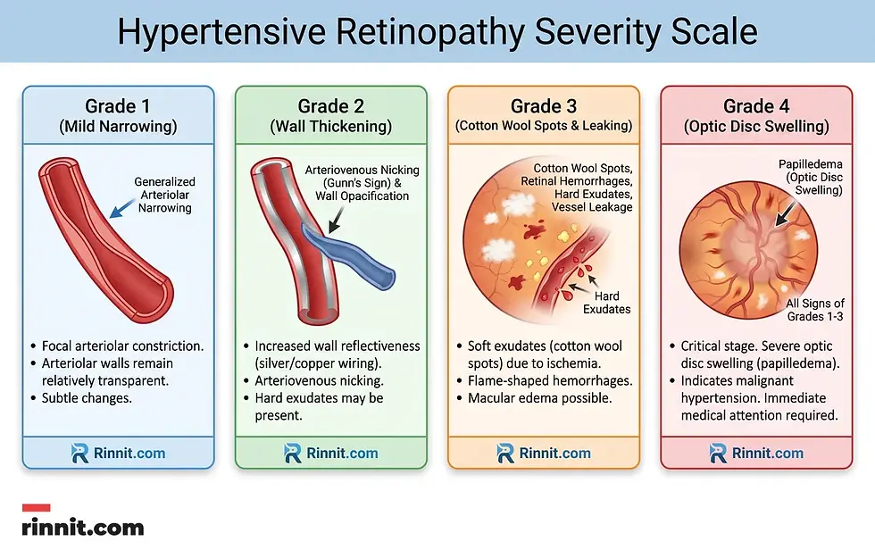
Healthcare providers generally use a four-grade system, or an alternative three-category system (mild, moderate, malignant), to describe severity, which helps determine the necessary level of intervention. [1]
Grade or category | Clinical observations |
Mild (Grade 1-2) | General narrowing of the arterioles and thickening of vessel walls. |
Moderate (Grade 3) | Presence of cotton wool spots (grayish-white clouds), microaneurysms, and hard exudates. |
Malignant (Grade 4) | Severe retinal damage including papilledema (optic disc swelling) and potential vision loss. |
Causes and systemic risk factors
The fundamental cause of hypertensive retinopathy is chronic, uncontrolled high blood pressure. Over time, the pressure causes retinal blood vessels to tighten (vasospasm) and eventually grow stiff and thick (arteriosclerosis). This process severely limits the amount of oxygen-rich blood reaching the retinal tissue. [1]
Several factors can increase an individual's likelihood of developing the condition: [1]
Cardiovascular history: A history of carotid artery disease or other heart-related issues.
Lifestyle factors: Smoking, tobacco use, and obesity significantly strain the vascular system.
Metabolic indicators: High cholesterol, prediabetes, and elevated creatinine levels (indicating kidney stress).
Demographics: The condition is more prevalent in adults over 40 and is more common among Black people and people of Chinese descent.
Complications of untreated damage
Without proper intervention to lower blood pressure, hypertensive retinopathy can lead to several serious eye complications. These include macular edema (swelling of the central retina), retinal detachment, and blockages in the arteries or veins that serve the eye (retinal artery or vein occlusion). In the most severe cases, the lack of blood flow to the retina and optic nerve can result in permanent blindness. [1]
Diagnosis and management strategies
Diagnosis is typically achieved through a comprehensive eye exam and advanced retinal imaging, such as optical coherence tomography (OCT). These tests allow eye care specialists to see micro-damage, such as tiny red dots (microaneurysms) or yellow spots (hard exudates) caused by leaking fats and proteins. [1]
The path to recovery
The primary goal of treatment is to bring blood pressure back to a healthy range. Research shows that some signs of hypertensive retinopathy go away over time with treatment, although this isn't always the case and depends on severity. Treatment started in the early stages gives the best chance of reversing retinal damage. [1]
Lifestyle adjustments: Maintaining a healthy weight, following a low-salt heart-healthy diet, and limiting alcohol intake are foundational steps.
Physical activity: Working up to at least 150 minutes of moderate-intensity exercise per week.
Medication adherence: Taking prescribed antihypertensive medications exactly as directed. It is critical never to stop these medications without a healthcare provider's approval. [1]
Protecting your eyes from high blood pressure

You may be able to prevent hypertensive retinopathy by seeing a healthcare provider each year for a checkup, having your blood pressure measured, and managing high blood pressure if your numbers are high. Regular eye exams matter too: most adults over 40 need a complete eye exam every year or two, and your eye care specialist can tell you how often you need one. [1]
Learn more. For more evidence-based health guides, visit Rinnit.com.
Frequently Asked Questions
Can I see the damage in the mirror?
No. The signs of hypertensive retinopathy, such as cotton wool spots or microaneurysms, are internal and can only be seen by a specialist using professional imaging tools.
Is vision loss from this condition permanent?
It depends on the severity. Early-stage damage can often be reversed with blood pressure control, but advanced damage like retinal detachment can lead to permanent vision loss.
How often should I get my eyes checked if I have high blood pressure?
Follow the schedule recommended by your eye care specialist. Most adults over 40 need a complete eye exam every year or two, and people with chronic hypertension may need them more often.
Does smoking really affect my eyes?
Yes. Smoking and tobacco use are recognized risk factors for hypertensive retinopathy because they damage blood vessels throughout the body, including those in the retina.
What are "cotton wool spots"?
Cotton wool spots are small, cloud-like grayish-white spots on the retina that indicate areas where the tissue is not getting enough blood flow.
Can children get hypertensive retinopathy?
While it is most common in adults over 40, anyone with severe, long-term high blood pressure can develop it, including children with underlying health issues.
Why is salt intake so important for my eyes?
Excess salt causes the body to retain fluid, which increases blood pressure. Lowering salt intake is one of the main ways to reduce the pressure on your retinal vessels.
Will my vision return to normal after treatment?
Some people have no vision loss and the signs go away once their blood pressure comes down. Others have vision loss or complications that need further treatment, so the extent of recovery depends on how much damage occurred before treatment started.
Is this the same as diabetic retinopathy?
No. While both involve damage to the retina's blood vessels, hypertensive retinopathy is caused by high blood pressure, while diabetic retinopathy is caused by high blood sugar. High blood pressure can also worsen diabetic retinopathy.
What is papilledema?
Papilledema is the swelling of the optic disc, which occurs in the most severe stages of hypertensive retinopathy and requires immediate medical attention.
Can high cholesterol make this condition worse?
High cholesterol is a recognized risk factor. It contributes to the stiffening and narrowing of blood vessels, which can further restrict blood flow.
Are there specific foods that help?
A heart-healthy diet like the Mediterranean diet, which is rich in fruits, vegetables, and lean proteins, supports overall vascular health, with a particular focus on lowering salt intake.
How long does it take for the retina to heal?
Healing varies by individual and severity. Some signs of damage go away over time with consistent blood pressure control, and your eye care specialist can monitor your progress.
Can I drive if I have this condition?
In early stages, vision is usually unaffected. However, if you have reached advanced stages with vision loss, you must consult your doctor about your ability to drive safely.
What happens if I stop my blood pressure medication?
Stopping medication can let your blood pressure rise again, which can worsen retinal damage and raise the risk of a stroke. Never stop taking blood pressure medication without your provider's approval.
Related Reading
Fluorescein Angiography: How the Eye Dye Test Works, How to Prepare and What Results Mean explains the eye dye test that shows changes in the retina's blood vessels.
References
Cleveland Clinic. Hypertensive Retinopathy: Causes, Symptoms & Treatment. Cleveland Clinic. Last updated June 18, 2023. https://my.clevelandclinic.org/health/diseases/25100-hypertensive-retinopathy
Liew G, Xie J, Nguyen H, et al. Hypertensive retinopathy and cardiovascular disease risk: 6 population-based cohorts meta-analysis. Int J Cardiol Cardiovasc Risk Prev. 2023;17:200180. PMID 36936860. https://doi.org/10.1016/j.ijcrp.2023.200180
Cheung CY, Biousse V, Keane PA, et al. Hypertensive eye disease. Nat Rev Dis Primers. 2022;8(1):14. PMID 35273180. https://doi.org/10.1038/s41572-022-00342-0
Dziedziak J, Zaleska-Żmijewska A, Szaflik JP, et al. Impact of Arterial Hypertension on the Eye: A Review of the Pathogenesis, Diagnostic Methods, and Treatment of Hypertensive Retinopathy. Med Sci Monit. 2022;28:e935135. PMID 35046380. https://doi.org/10.12659/MSM.935135
Medical disclaimer
This article is for general educational purposes only and is not a substitute for professional medical advice, diagnosis, or treatment. If you have high blood pressure or are experiencing vision changes, consult a healthcare professional.



Comments