Fluorescein Angiography: How the Eye Dye Test Works, How to Prepare and What Results Mean

Updated: 2 days ago
Written and medically reviewed by Dr. Baraa Alnahhal, MD · Last reviewed: November 2026 · How we review
Quick Answer: What is fluorescein angiography?
Fluorescein angiography is an eye imaging test that photographs the blood vessels of the retina. An eye specialist widens your pupils with drops, injects a fluorescent dye into a vein and takes pictures as the dye flows through the eye. It takes less than 30 minutes, is low risk and helps diagnose diabetic retinopathy and macular degeneration. [1]
What the research says
Serious reactions to the dye are rare. A review of 78 studies published over more than 50 years found overall reaction rates of 0.083% to 21.69%, but severe reactions in only 0.04% to 0.59% of tests; the only proven way to lower risk was checking for a past reaction to fluorescein. (Kornblau and El-Annan, Survey of Ophthalmology, 2019) [2]
A past reaction is the strongest warning sign. In 3,381 consecutive tests in 2,247 patients, 3.3% had a side effect, and people who had reacted before were 6.2 times more likely to react again. (Xu et al., Canadian Journal of Ophthalmology, 2016) [3]
Wide-view cameras see much more of the retina. Ultra-widefield imaging can capture about 82% of the retina, compared with about 30% for standard photography, and in diabetes eyes with mostly peripheral changes progressed faster. (Ashraf et al., Seminars in Ophthalmology, 2020) [4]
Key takeaways
Fluorescein angiography uses an injected dye and a special camera to show blood flow in the retina. [1]
It helps find leaks or blockages linked to diabetic retinopathy, macular degeneration and other retinal conditions. [1]
Dilating drops can blur your vision for up to 12 hours, so arrange a ride home. [1]
Yellowish skin and dark yellow or orange urine are common and harmless after the test. [1]
Allergic reactions are rare, but breathing trouble after the dye is an emergency. [1]
What is fluorescein angiography?
Fluorescein angiography is an imaging test that takes photographs of the blood vessels inside your retina. The retina is the thin, light-sensitive tissue at the back of your eye that turns light into signals and sends them to your brain. [1]
The test relies on two things: eye drops that dilate (widen) your pupils, and a fluorescent dye that travels through your bloodstream. When the dye reaches your eye, it makes the retinal blood vessels glow so the camera can capture them clearly. An ophthalmologist, a doctor who specializes in eye disease, performs and reads the test. [1]
Why is fluorescein angiography done?
Eye specialists use the test to diagnose retinal conditions and to track how they change over time or respond to treatment. Conditions it can help with include: [1]
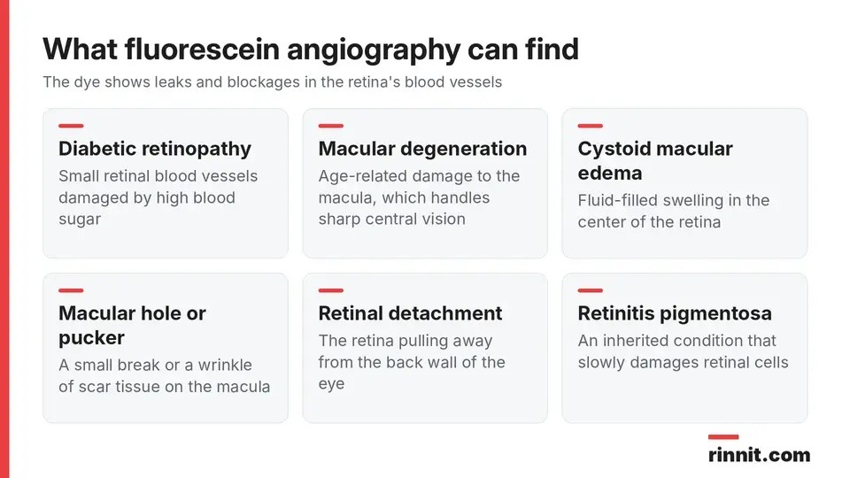
The dye test shows leaks and blockages in retinal blood vessels.
Condition | What it affects |
Age-related macular degeneration | The macula, the center of the retina that handles sharp central vision |
Diabetic retinopathy | Small retinal blood vessels damaged by high blood sugar |
Cystoid macular edema | Fluid-filled swelling in the macula |
Macular hole or macular pucker | A small break or a wrinkle of scar tissue on the macula |
Retinal detachment | The retina pulling away from the back wall of the eye |
Retinitis pigmentosa | An inherited condition that slowly damages retinal cells |
Ocular melanoma | A cancer that can develop inside the eye |
Sources: [1]
How do I prepare for fluorescein angiography?
A few simple steps help the test go smoothly: [1]
Arrange a ride: dilating drops can blur your vision for up to 12 hours, so you should not drive yourself home
Review your medicines: tell your provider about everything you take, since some medicines may need to be paused for a short time
Mention allergies: let your provider know about any allergies, especially to iodine
Tell them about pregnancy: say if you are or might be pregnant, because the effect of the test on a developing baby is not known
Skip contact lenses: take your contacts out on the day of the test
What happens during fluorescein angiography?
The test usually takes place in your eye doctor's office and lasts less than 30 minutes. [1]

The whole test usually takes less than 30 minutes.
Dilation: you get eye drops to widen your pupils
Positioning: you rest your chin and forehead against the camera and keep your head still
First photos: the camera takes a set of baseline pictures of your retina
Dye injection: a provider injects the fluorescent dye into a vein in your arm
More photos: the camera takes more pictures as the dye moves through the blood vessels in your eye
The test should not hurt. If you feel pain at any point, tell your provider right away. [1]
What should I expect after the test?
You can go home the same day, but someone else should drive you. Light sensitivity and blurry vision are common and can last for several hours, so wearing sunglasses helps until your eyes recover. Many people take the rest of the day off work while their pupils return to their normal size. [1]
What are the risks and side effects of fluorescein angiography?
Fluorescein angiography is generally a low-risk test. Most side effects are short-lived and harmless: [1]

Most side effects fade within minutes to a day.
Side effect | How long it usually lasts |
Dark or tinted vision | Several minutes |
Blurry vision and light sensitivity from dilation | Several hours (up to 12 hours) |
Slight yellowing of the skin | Up to several hours |
Dark yellow or orange urine | Up to 24 hours |
Burning at the injection site if dye leaks under the skin | Brief; it does not harm the skin |
Sources: [1]
Some people also notice a short-lived metallic taste, dry mouth, dizziness, a faster heartbeat, sneezing, nausea or vomiting. Rarely, the dye causes an allergic reaction with itching or hives. [1]
Severe allergic reactions (anaphylaxis) are very rare but can be life-threatening. In large studies, serious reactions occurred in well under 1% of tests, and a previous reaction to the dye was the clearest risk factor. [2] [3]
What do fluorescein angiography results mean?
Your ophthalmologist usually goes over your results within a few days. [1]
If the photos show dye leaking from blood vessels or vessels that look blocked, that pattern can point to conditions such as diabetic retinopathy or macular degeneration. Your provider may order more tests to confirm the cause and plan treatment. [1]
When should I call my provider or get emergency care?
Contact your provider if you notice signs of an allergic reaction, such as itching or hives, after the test. Call 911 or go to the nearest emergency room if you have any trouble breathing, because it can be a sign of a severe reaction. [1]
Conclusion
Fluorescein angiography gives your eye specialist a detailed map of the blood vessels in your retina, which helps them find leaks or blockages that a routine exam might miss. The test is quick and low risk, and its most common effects, such as blurry vision and yellow-tinted skin or urine, fade within a day.
Plan ahead with a driver and sunglasses, share your medicines and allergies, and mention any past reaction to the dye so your care team can keep the test as safe as possible.
Keep learning: Read our eye exam guide or our glaucoma tests guide. For more background, see MedlinePlus on fluorescein angiography.
FAQ
What is fluorescein angiography?
An eye test that uses an injected dye and a camera to photograph the blood vessels in your retina.
Why would I need fluorescein angiography?
To diagnose or monitor retinal conditions such as diabetic retinopathy or macular degeneration.
How long does fluorescein angiography take?
Usually less than 30 minutes in your eye doctor's office.
Does fluorescein angiography hurt?
It should not be painful, though you may feel the needle or a brief burn if dye leaks.
Can I drive after fluorescein angiography?
No. Dilating drops can blur your vision for up to 12 hours, so arrange a ride.
Why is my urine orange after fluorescein angiography?
The dye leaves your body through your urine, which can look dark yellow or orange for up to 24 hours.
Why does my skin look yellow after the test?
The dye can tint your skin slightly yellow for up to several hours; this is harmless.
Is fluorescein angiography safe?
Yes, it is low risk; serious allergic reactions are rare.
Can I have fluorescein angiography while pregnant?
Tell your provider if you are or may be pregnant, since the effect on a developing baby is not known.
When will I get my results?
Your eye doctor usually discusses them within a few days.
Related Reading
Diabetic Retinopathy: Symptoms, Causes, Diagnosis & Treatment — Complete Guide explains one of the most common reasons for this dye test.
Dry Macular Degeneration: Complete Guide to Symptoms, Causes & Treatment covers a macular condition that retinal imaging helps monitor.
Retinal Vein Occlusion (RVO): Symptoms, Types, and Treatment describes a blocked retinal vein, a problem angiography can show.
Uveitis: Symptoms, Causes, and Treatment — What Eye Inflammation Means for Your Vision explains eye inflammation that can make retinal vessels leak.
Central Serous Retinopathy: The "Stress Eye Disease" That Can Blur Your Vision — and Usually Goes Away on Its Own covers fluid under the retina that eye specialists may assess with dye imaging.
Hypertensive Retinopathy: A Comprehensive Guide to Blood Pressure-Related Eye Damage explains how high blood pressure damages the retinal blood vessels this test photographs.
References
Cleveland Clinic. Fluorescein Angiography: What It Is and What To Expect. Cleveland Clinic. Last updated December 27, 2022. https://my.clevelandclinic.org/health/diagnostics/24618-fluorescein-angiography
Kornblau IS, El-Annan JF. Adverse reactions to fluorescein angiography: A comprehensive review of the literature. Surv Ophthalmol. 2019;64(5):679-693. PMID 30772364. https://doi.org/10.1016/j.survophthal.2019.02.004
Xu K, Tzankova V, Li C, et al. Intravenous fluorescein angiography-associated adverse reactions. Can J Ophthalmol. 2016;51(5):321-325. PMID 27769320. https://doi.org/10.1016/j.jcjo.2016.03.015
Ashraf M, Shokrollahi S, Salongcay RP, et al. Diabetic retinopathy and ultrawide field imaging. Semin Ophthalmol. 2020;35(1):56-65. PMID 32167854. https://doi.org/10.1080/08820538.2020.1729818
Medical disclaimer
This article is for general education only and does not replace advice from a qualified healthcare professional. Talk to your doctor about your own diagnosis and treatment, and seek emergency care right away for severe or sudden symptoms.



Comments