Retinal Vein Occlusion (RVO): Symptoms, Types, and Treatment

Updated: 2 hours ago
Medically reviewed by Dr. Baraa Alnahhal, MD · Last reviewed: September 2026
TL;DR
Retinal Vein Occlusion (RVO) is a blockage in the veins that drain blood from the retina. It is a common cause of vision loss, typically affecting one eye with symptoms like sudden blurring or floaters. While the blockage itself cannot be reversed, treatments such as anti-VEGF injections and laser therapy are highly effective at managing complications like swelling and abnormal blood vessel growth.
Quick Answer
What is Retinal Vein Occlusion (RVO)?
RVO is a vascular disorder where a blood clot or vessel compression blocks blood flow out of the retina. This leads to increased pressure, swelling, and potential vision loss. It is the second most common retinal disorder after diabetic retinopathy. Prompt diagnosis and treatment of complications are essential to prevent permanent eye damage and preserve sight.
WARNING: SUDDEN VISION CHANGES REQUIRE URGENT CARE While RVO is often painless, it can lead to severe complications. Seek immediate medical evaluation if you experience any of the following:
Sudden or rapid loss of vision in one eye.
A dramatic increase in floaters or dark spots.
Severe eye pain or a feeling of intense pressure.
Sudden, unexplained blurring of your sight.

What is Retinal Vein Occlusion?
Retinal Vein Occlusion (RVO) occurs when one of the veins responsible for carrying blood away from the retina becomes partially or totally blocked. The retina is the light-sensitive tissue at the back of the eye that converts light into the signals your brain uses to create images. When a vein is blocked, blood cannot leave the eye properly, leading to a backup of fluid, increased intraocular pressure, and swelling.
There are two primary forms of this condition:
Central Retinal Vein Occlusion (CRVO): A blockage in the main vein that drains the entire retina.
Branch Retinal Vein Occlusion (BRVO): A blockage in one of the smaller veins branching off the main vessel. BRVO is the more common type.
Symptoms and Warning Signs
RVO typically affects only one eye. The symptoms can vary depending on the severity of the blockage and whether complications have already developed.
Blurry Vision: This is the most common symptom and may occur suddenly or develop over several days.
Vision Loss: A partial or complete loss of sight in the affected eye.
Eye Floaters: The appearance of dark spots, lines, or "cobwebs" in your field of vision.
Eye Pain or Pressure: Typically seen in advanced cases where internal eye pressure has risen significantly.
Causes and Risk Factors
RVO is caused by a disruption in normal blood flow. This can happen if a blood clot forms in the vein or if a nearby artery, stiffened by age or plaque, compresses the vein.
Risk Factor | Why it Matters |
|---|---|
Age | Most cases occur in people over 50, though it can affect younger adults. |
High Blood Pressure | Hypertension is a leading contributor to vessel damage and compression. |
Diabetes | High blood sugar can damage the delicate blood vessels in the eye. |
Atherosclerosis | Hardening of the arteries increases the risk of vein compression. |
Glaucoma | Increased eye pressure can contribute to vascular blockages. |
Potential Complications
If left untreated, RVO can lead to several serious eye conditions:
Macular Edema: Swelling in the center of the retina (the macula), causing blurred central vision.
Neovascularization: The growth of abnormal, fragile blood vessels that are prone to leaking.
Vitreous Hemorrhage: Bleeding into the gel-like substance that fills the eye.
Neovascular Glaucoma: A painful increase in eye pressure caused by abnormal vessel growth.
Retinal Detachment: In severe cases, abnormal vessels can pull the retina away from the back of the eye.
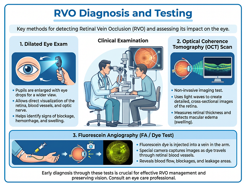
Diagnosis and Testing
Eye care specialists use several advanced imaging tools to diagnose RVO and assess the extent of the damage.
Dilated Eye Exam: Using drops to widen the pupil, the doctor examines the retina for signs of blockage or bleeding.
Optical Coherence Tomography (OCT): A high-resolution scan that measures retinal thickness and identifies macular edema.
Fluorescein Angiography: A dye is injected into the arm to highlight the blood vessels in the retina, showing exactly where the blockage is located.
Fundus Photography: Detailed photos of the back of the eye to document abnormal vessel growth and bleeding.
Treatment and Management
While the original blockage usually cannot be cleared, treatments focus on managing complications and protecting your remaining vision.
Medical Injections
Anti-VEGF Injections: These medications block a protein (VEGF) that causes abnormal blood vessel growth and leakage. This is the primary treatment for macular edema.
Steroid Injections: Used to reduce inflammation and swelling in the retina.
Surgical and Laser Procedures
Panretinal Photocoagulation (PRP): A laser treatment used to shrink abnormal blood vessels and prevent further bleeding or pressure buildup.
Vitrectomy: A surgery to remove blood or the gel-like vitreous from the eye if a hemorrhage has occurred.

Conclusion
Retinal Vein Occlusion is a serious vascular condition that can significantly impact your vision. While the blockage itself is often permanent, modern medical interventions are highly successful at preventing further damage and managing complications like swelling and bleeding. Early detection through regular eye exams is the most effective way to protect your sight.
Next Steps
If you notice any sudden changes in your vision, see an eye specialist immediately. Managing underlying health conditions like high blood pressure and diabetes is critical for preventing RVO in your other eye and reducing the risk of systemic issues like stroke.
Frequently Asked Questions
1. Can Retinal Vein Occlusion be cured?
There is no current way to unblock the vein, but the complications (like swelling and bleeding) can be treated effectively to save your vision.
2. Is RVO a sign of a stroke?
While RVO is a blockage in the eye, people with RVO have a higher risk of cardiovascular issues, including stroke, due to shared risk factors like high blood pressure.
3. Does RVO affect both eyes?
It usually affects only one eye at a time, but having it in one eye increases the risk of it developing in the other eye later.
4. What is the difference between CRVO and BRVO?
CRVO is a blockage of the main retinal vein, while BRVO is a blockage of a smaller branch vein. BRVO is generally more common.
5. How long do the injections last?
Anti-VEGF injections are often given in a series, sometimes monthly, depending on how your eye responds to the treatment.
6. Will my vision ever return to normal?
Many patients experience significant improvement with treatment, but the final outcome depends on the severity of the blockage and how quickly treatment began.
7. Can I prevent RVO?
You can lower your risk by managing high blood pressure, cholesterol, and diabetes, and by not smoking.
8. Is laser treatment painful?
Most patients experience only mild discomfort during laser procedures. Your doctor will use numbing drops or local anesthesia.
9. What are floaters?
Floaters are small spots or lines in your vision caused by clumps in the eye's gel or bleeding from abnormal vessels.
10. Can high eye pressure cause RVO?
Yes, conditions like glaucoma that increase internal eye pressure are known risk factors for vein occlusion.
11. What is macular edema?
It is the buildup of fluid and swelling in the macula, the part of the retina responsible for sharp, central vision. Read more in our guide to cystoid macular edema.
12. How is RVO diagnosed?
It is diagnosed through a dilated eye exam and imaging tests like OCT and fluorescein angiography.
13. What happens if RVO is left untreated?
It can lead to permanent vision loss, neovascular glaucoma, and retinal detachment.
14. Are there side effects to the injections?
Common side effects include temporary eye irritation or a feeling of something in the eye. Serious complications like infection are very rare.
15. Should I see my regular doctor too?
Yes. Your eye specialist will likely coordinate with your primary care physician to manage underlying risks like hypertension.
Related Reading
Trusted External Resources
References
Medical Disclaimer
This article is for informational purposes only and does not constitute medical advice. Always seek the advice of your physician or other qualified health provider with any questions you may have regarding a medical condition. Never disregard professional medical advice or delay in seeking it because of something you have read in this article.
Comments