Papilledema: Causes, Symptoms, and Emergency Care

Updated: 3 days ago
Medically reviewed by Dr. Baraa Alnahhal, MD · Last reviewed: October 2026
Papilledema is swelling of the optic discs in both eyes, caused by increased pressure inside the skull. It can be a medical emergency because the pressure may have a serious cause, and untreated papilledema can lead to partial or complete blindness [1].
Quick Answer: What is papilledema?
Papilledema is swelling of the optic discs caused by raised pressure around the brain, called intracranial hypertension. Symptoms include headaches worse in the morning or when lying down, brief vision blackouts, double vision and nausea. Diagnosis involves an eye exam, MRI and sometimes a lumbar puncture, and treatment targets the cause of the pressure. [1]
What the research says
Acetazolamide plus weight loss modestly reduces optic disc swelling in IIH. A Cochrane review of two randomized trials (211 participants) found that in one study the acetazolamide group had a greater reduction in papilloedema by clinical grading over six months (mean difference −0.91, 95% CI −1.27 to −0.54). The authors rated the evidence as low certainty and called for more high-quality trials (Piper et al., Cochrane Database Syst Rev, 2015).
Venous sinus stenting helps some people who do not respond to medicine. A meta-analysis of 36 studies and 1,066 patients with idiopathic intracranial hypertension found papilledema improved in 89% after stenting, while 13.7% had treatment failure or complications (Azzam et al., J Neurol Sci, 2024).
AI is getting good at spotting disc swelling on photos. A systematic review of 32 studies found deep learning models detected papilledema on fundus photographs with AUC above 0.98, but performance was only modest at telling mild papilledema from pseudopapilledema on OCT scans (Fang and Chen, BMC Ophthalmol, 2026).
Research summaries are based on article abstracts retrieved from PubMed.
What Is Papilledema?
The optic disc, also called the nerve head, is where the optic nerve enters the back of the eye. The optic nerve is the pathway that connects the eye to the brain. Cerebrospinal fluid is constantly released and reabsorbed, and normal levels help protect the brain from trauma. When pressure rises, both optic discs can swell [1].
Papilledema almost always affects both eyes, because it is driven by pressure inside the skull. Swelling of the optic disc in only one eye is usually not caused by intracranial hypertension and is called optic disc edema [1]. Pseudopapilledema is different again: the optic disc looks raised, but it is not because of optic nerve swelling [1].

Anatomical cross-section of the eye showing a swollen optic disc and the impact of increased intracranial pressure.
What are the symptoms of papilledema?
You may have no symptoms, especially early on. When symptoms do occur, they can change with posture and time of day [1].
Symptom | What to know |
|---|---|
Headaches | Often worse in the morning and when you are lying down |
Transient visual obscurations | Episodes of about 5 to 15 seconds when vision blurs, goes gray, or blacks out, usually when you change posture |
Double vision (diplopia) | Can happen if raised pressure affects the cranial nerves that control the eye muscles |
Nausea and vomiting | Can occur along with the other symptoms of raised pressure |
Neurological symptoms | May include problems with movement or thinking |
Source: Cleveland Clinic [1].
Vision loss worsens as the condition progresses. Without treatment, papilledema can lead to partial or complete blindness in one or both eyes [1].
What causes papilledema and who is at risk?
Papilledema is caused by high intracranial pressure, so there is always an underlying issue to find and treat [1].
Who is most affected?
Papilledema is more common in women, usually aged 20 to 44, who have overweight (a BMI greater than 25) or obesity (a BMI greater than 30). In this group the incidence is 13 per 100,000, compared with 0.9 per 100,000 in the U.S. population as a whole [1].
Related causes
Idiopathic intracranial hypertension (IIH): raised pressure with no clear, identifiable cause
Malignant hypertension: dangerously high blood pressure
Tumors
Infection, bleeding, or inflammation in the brain or the meninges, the tissues that protect the brain and spinal cord
Cerebral venous sinus thrombosis: a blood clot in a vein in the brain
Iron-deficiency anemia
Medications and other triggers: links have been reported with retinoids, toxic amounts of vitamin A, tetracycline antibiotics, corticosteroids, and COVID-19
Source: Cleveland Clinic [1].
How is papilledema diagnosed?
Papilledema is often first found during an evaluation for headaches or vision problems, and diagnosis usually involves an eye exam, imaging, a lumbar puncture and blood tests. Because it can be life-threatening, the work-up is often urgent. The usual steps are [1]:
Eye exam: an eye doctor looks at the optic disc for swelling and may add visual field testing.
Imaging: if the disc looks swollen, an MRI scan is the most likely next test.
Lumbar puncture (spinal tap): your provider may order one to measure the pressure of the cerebrospinal fluid.
Blood tests: these check iron and other substances in the blood.
Providers may grade severity with a version of the Frisén scale, which runs from Grade 0 (a normal disc, though the edges may be blurred) to Grade V (the whole disc is elevated and the blood vessels in and leaving the disc cannot be seen) [1]. Researchers are also testing artificial intelligence to help detect disc swelling on photos and OCT scans, but these tools are still early-stage and do not replace a specialist exam [4].

Visual guide to the diagnostic pathway for papilledema, including ophthalmoscopy, MRI, and lumbar puncture.
How is papilledema treated?
The goal is to lower intracranial pressure and protect the optic nerve, so treatment depends on the cause [1]:
Idiopathic intracranial hypertension: a carbonic anhydrase inhibitor such as acetazolamide, together with achieving and maintaining a healthy weight as a long-term strategy.
If those measures are not enough: surgical procedures to relieve and decrease the pressure.
Other causes: your provider treats the underlying cause, such as a tumor, infection, blood clot, or high blood pressure.
In trials of acetazolamide plus weight loss for IIH, optic disc swelling improved modestly, although the overall evidence was rated low certainty [2]. For selected people who do not respond to medicine, venous sinus stenting has been reported to improve papilledema in most patients, with treatment failure or complications in a minority [3].
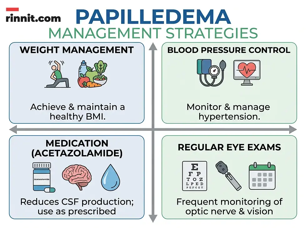
Infographic detailing management strategies for papilledema, including weight management, medication, and blood pressure control.
Can papilledema be prevented, and what is the outlook?
If papilledema is caught early, the outlook is good, because treatment mainly addresses the issue that caused it. You can lower your risk by keeping your blood pressure under control and by achieving and keeping a healthy weight. Regular eye exams matter too, since early diagnosis leads to better outcomes [1].
When should you see a doctor about papilledema?
Seek immediate medical care if you have new or worsening headaches along with nausea and vomiting. Have any change in your vision assessed by a healthcare provider, particularly when it comes with headaches, nausea, or vomiting [1]. If you notice new morning headaches or brief vision blackouts, see an eye specialist or emergency care provider promptly so your intracranial pressure can be checked.
What is the bottom line on papilledema?
Papilledema is swelling of both optic discs caused by raised pressure in the skull, and it can signal a serious problem. Morning headaches and brief vision blackouts are the classic warning signs. Finding and treating the underlying cause, whether with medication, weight management, or surgery, gives most people the best chance of protecting their vision [1].
Frequently Asked Questions
What is papilledema?
Papilledema is swelling of both optic discs, where the optic nerves enter the back of the eyes, caused by increased intracranial pressure.
Is papilledema a medical emergency?
Papilledema can be. It points to high pressure in the brain, which may come from serious causes such as tumors, infection, or blood clots.
Can papilledema cause permanent blindness?
Yes. Untreated papilledema can lead to partial or complete blindness in one or both eyes.
Does papilledema affect both eyes?
Almost always. Swelling in only one eye is usually not due to intracranial hypertension and is called optic disc edema.
What is the difference between papilledema and pseudopapilledema?
In pseudopapilledema the optic disc looks raised but is not swollen from optic nerve swelling or brain pressure.
How long do the vision blackouts last?
Transient visual obscurations last about 5 to 15 seconds and usually happen when you change posture.
Can weight loss help treat papilledema?
For papilledema caused by idiopathic intracranial hypertension, reaching and keeping a healthy weight is a long-term strategy.
Do migraines cause papilledema?
No. Migraines do not cause papilledema, although both can cause severe headaches.
How is papilledema graded?
Providers may use a version of the Frisén scale, from Grade 0 (normal disc) to Grade V (whole disc elevated with vessels obscured).
Is papilledema hereditary?
No. Papilledema is not hereditary.
Can anemia cause papilledema?
Iron-deficiency anemia is listed among the conditions related to raised intracranial pressure and papilledema.
Related Resources
External resource for deeper reading:
Cleveland Clinic. Papilledema. Cleveland Clinic. Medically reviewed; last updated November 13, 2022. https://my.clevelandclinic.org/health/diseases/24445-papilledema
Related Reading
Fundoscopy (Ophthalmoscopy): What It Is, Types, What to Expect and What It Can Find explains the exam of the retina and optic nerve used in eye and diabetes care.
Fundus Photography: What It Shows, What to Expect and Who Needs It explains the eye photos used to check the retina and optic nerve.
References
Cleveland Clinic. Papilledema. Cleveland Clinic Health Library. Last updated November 13, 2022. https://my.clevelandclinic.org/health/diseases/24445-papilledema
Piper RJ, Kalyvas AV, Young AM, et al. Interventions for idiopathic intracranial hypertension. Cochrane Database Syst Rev. 2015;2015(8):CD003434. PMID: 26250102. DOI: 10.1002/14651858.CD003434.pub3
Azzam AY, Mortezaei A, Morsy MM, et al. Venous sinus stenting for idiopathic intracranial hypertension: an updated meta-analysis. J Neurol Sci. 2024;459:122948. PMID: 38457956. DOI: 10.1016/j.jns.2024.122948
Fang SS, Chen SH. AI-assisted diagnosis of neuro-ophthalmic disorders: a systematic review from optic neuritis to papilledema. BMC Ophthalmol. 2026;26(1):274. PMID: 41947081. DOI: 10.1186/s12886-026-04772-2
Medical disclaimer
This article is for informational purposes only and is not a substitute for professional medical advice, diagnosis, or treatment. Always seek the advice of your physician or other qualified health provider with any questions you may have about a medical condition.



Comments