Fundus Photography: What It Shows, What to Expect and Who Needs It

Updated: 2 days ago
Written and medically reviewed by Dr. Baraa Alnahhal, MD · Last reviewed: November 2026 · How we review
Quick Answer
Fundus photography is a quick, painless test that takes detailed pictures of the back inside wall of the eye, the fundus, which includes the retina, macula, optic nerve and blood vessels. The photos help detect and monitor diabetic retinopathy, macular degeneration and glaucoma, sometimes before you notice symptoms. You may get dilating drops, which cause brief blurry vision.
What the research says
Fundus photos can be read accurately, even by computers: an artificial intelligence system trained on retinal fundus photographs detected diabetic retinopathy needing referral with about 97.5% sensitivity and 93.4% specificity in a large validation set, showing how much these images reveal (Gulshan V et al., JAMA, 2016) [2]
A single photo can outperform a quick eye check: in 197 people with diabetes, a single nondilated digital fundus photograph read remotely detected diabetic retinopathy with 78% sensitivity, compared with just 34% for dilated ophthalmoscopy, when both were checked against a standard set of seven photographs (Lin DY et al., American Journal of Ophthalmology, 2002) [3]
Key takeaways
Photographs the retina, macula, optic nerve and blood vessels at the back of the eye.
Helps find diabetic retinopathy, macular degeneration, glaucoma, retinal tears and more.
Quick and painless; dilating drops may be used.
Wide-field cameras can often take photos without dilation.
Photos create a record to compare changes over time.
What is the fundus?
The fundus is the inside back surface of the eye. It includes the retina, the light-sensing layer; the macula, which gives sharp central vision; the optic nerve; and the blood vessels and choroid that nourish them. Because it's the only place where doctors can directly see blood vessels and nerve tissue, it can also show signs of diabetes, high blood pressure and brain swelling. Sources: [1]
Why is it done?

Fundus photos show the retina, macula and optic nerve.
Diabetic retinopathy and high blood pressure damage to the retina.
Glaucoma damage to the optic nerve.
Optic nerve problems such as optic atrophy and papilledema.
Uveitis, blind spots, eye tumors such as retinoblastoma, and infections such as toxoplasmosis. Sources: [1]
It's often part of a routine eye exam and may be combined with optical coherence tomography (OCT) or fluorescein angiography. Unlike fundoscopy, which is a live look by your provider, photography creates a permanent record. Sources: [1]
Types of fundus cameras

Wide-field cameras can often work without dilation.
Camera | How it works | Dilation |
Standard fundus camera | Telescope-like camera with chin and forehead rests; photos taken one at a time | Usually works best with dilating drops |
Wide-field laser camera | Scans a wide area of the retina in about a second | Often not needed |
Sources: [1]
How do I prepare?
No preparation is needed. If you'll get dilating drops, bring sunglasses and consider having someone drive you, since your vision may be blurry and light-sensitive for a few hours. Sources: [1]
What happens during the test?
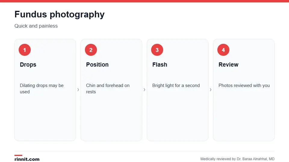
Fundus photography is quick and painless.
You may get dilating eye drops, which take several minutes to work.
You sit and rest your chin and forehead on the camera supports.
You look at a target light while the specialist lines up the camera.
A bright flash or a quick sweep of light takes each photo; several are usually taken per eye. Sources: [1]
Risks
Fundus photography is very safe and has no side effects. The flash may feel bright for a moment. Dilating drops cause blurry vision and light sensitivity for a few hours; disposable sunglasses are often provided. Sources: [1]
Understanding your results
Photos are often reviewed with you right away on a screen. If everything looks normal, no further action is needed. If changes are found, your specialist may compare them with earlier photos and recommend more tests, treatment, follow-up or a referral. Sources: [1, 2, 3]
Conclusion
Fundus photography is a fast, painless way to capture the health of the back of your eye. For people with diabetes, high blood pressure or glaucoma, regular photos help catch changes early, often before vision is affected.
Keep learning: read about diabetic retinopathy and the eye exam.
Frequently asked questions
What is fundus photography?
Taking pictures of the back inside wall of the eye.
What does a fundus photo show?
The retina, macula, optic nerve and blood vessels.
Does it hurt?
No. The flash is bright but painless.
Will my eyes be dilated?
Sometimes; wide-field cameras often don't need dilation.
Can I drive afterward?
If your eyes were dilated, it's best to have someone else drive.
How long does it take?
Usually just a few minutes, plus time for drops to work.
Why is it done for diabetes?
To find and track diabetic retinopathy early.
Is it the same as OCT?
No. OCT takes cross-section images; fundus photos show the surface.
Is it the same as fundoscopy?
No. Fundoscopy is a live look; photography makes a record.
Are there risks?
No significant risks; drops cause temporary blurry vision.
Related Reading
Diabetic Retinopathy: Symptoms, Causes, Diagnosis & Treatment — Complete Guide diabetes damage to the retina found on photos.
Glaucoma: The Silent Threat to Vision, and How Early Treatment Can Protect Your Sight optic nerve damage tracked with fundus photos.
Dry Macular Degeneration: Complete Guide to Symptoms, Causes & Treatment central vision loss seen in the macula.
Retinal Detachment: Symptoms, Causes, Diagnosis & Treatment — Complete Guide a sight-threatening emergency.
Papilledema: Causes, Symptoms, and Emergency Care optic nerve swelling from brain pressure.
Retinal Imaging: Fundus Photos, OCT and Angiography Explained explains the photos and scans used to check the back of the eye.
References
Cleveland Clinic. Fundus Photography: What It Is & When It Is Needed. Cleveland Clinic. Last updated July 23, 2024. https://my.clevelandclinic.org/health/diagnostics/fundus-photography
Gulshan V, Peng L, Coram M, et al. Development and validation of a deep learning algorithm for detection of diabetic retinopathy in retinal fundus photographs. JAMA. 2016;316(22):2402-2410. PMID 27898976. https://doi.org/10.1001/jama.2016.17216
Lin DY, Blumenkranz MS, Brothers RJ, et al. The sensitivity and specificity of single-field nonmydriatic monochromatic digital fundus photography with remote image interpretation for diabetic retinopathy screening: a comparison with ophthalmoscopy and standardized mydriatic color photography. Am J Ophthalmol. 2002;134(2):204-213. PMID 12140027. https://doi.org/10.1016/s0002-9394(02)01522-2
Medical disclaimer
This article is for general education only and does not replace advice from a qualified healthcare professional. Talk to your doctor about your own diagnosis and treatment, and seek emergency care right away for severe or sudden symptoms.



Comments