Optical Coherence Tomography (OCT) Eye Test: What It Shows and What to Expect

Updated: 2 days ago
Written and medically reviewed by Dr. Baraa Alnahhal, MD · Last reviewed: November 2026 · How we review
Quick Answer: What is an OCT eye test?
Optical coherence tomography (OCT) is a quick, noninvasive scan that uses reflected infrared light to create cross-section images of the back of the eye, including the retina and optic nerve. It helps diagnose and track glaucoma, diabetes-related retinopathy and macular degeneration. The scan takes a few minutes, nothing touches the eye, and it has no known risks. [1]
What the research says
OCT helps detect glaucoma but works best alongside other tests. A Cochrane review of 106 studies (16,260 eyes) found that average nerve fiber layer thickness on OCT had about 72% sensitivity and 93% specificity for manifest glaucoma, similar to other imaging devices. (Michelessi et al., Cochrane Database of Systematic Reviews, 2015) [2]
OCT is now the reference test for diabetic macular swelling. In 9 studies (759 people, 1,303 eyes), OCT detected clinically significant diabetic macular edema with 78% sensitivity and 86% specificity, and the authors concluded OCT is widely recognized as the new reference standard. (Virgili et al., Cochrane Database of Systematic Reviews, 2015) [3]
Key takeaways
OCT makes layered, cross-section images of the retina and optic nerve using light, not radiation. [1]
It helps diagnose and monitor glaucoma, diabetic eye disease, macular degeneration and retinal problems. [1]
You don't need to prepare, and the scan takes only a few minutes. [1]
Your pupils may be dilated first, which can cause light sensitivity and blurry vision for a few hours. [1]
Repeat scans let your provider compare changes in your eye over time. [1]
What is optical coherence tomography?
OCT is a noninvasive imaging method that eye care specialists use to see cross-sections of your eye. It measures infrared light that bounces off your retina, letting providers look at the eye layer by layer and measure the thickness of key structures. [1]
When it's used on the eye, it's sometimes called ocular coherence tomography. Providers in cardiology, neurology and oncology now use OCT too, including for imaging blood vessels (angiography). [1]
How does OCT work?
OCT works much like ultrasound, another imaging test that doesn't touch the area being studied. Ultrasound bounces sound waves off tissue, while OCT bounces invisible light off your tissues and measures what returns, building three-dimensional cross-section images. [1]
What can an OCT scan help diagnose?
OCT is most often used to look at your retina and optic nerve. It helps diagnose retinal diseases and related conditions that can affect your vision, including: [1]
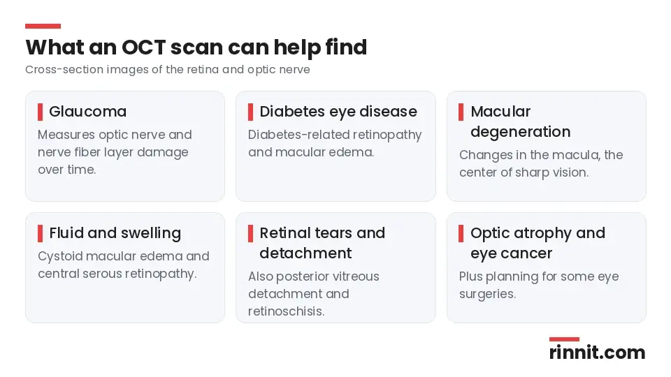
Area | Conditions |
Optic nerve | Glaucoma, optic atrophy |
Macula | Macular degeneration, cystoid macular edema, diabetes-related macular edema, bull's eye maculopathy |
Retina | Diabetes-related retinopathy, central serous retinopathy, retinal tears, retinal detachment, retinoschisis |
Other | Posterior vitreous detachment, eye cancer |
Sources: [1]
OCT can also image the front of your eye to help find defects there or plan eye surgery. [1]
When should you have an OCT scan?
Your provider may suggest OCT if you have symptoms of an eye condition or if they see signs of one during an eye exam. If you're at risk of age-related eye disease or other eye problems, OCT may become part of your regular eye exams so scans can be compared over time. [1]
How do you prepare?
You don't need to do anything to prepare. The scan can even be done right after a standard eye exam. If your pupils will be dilated, plan for someone else to drive you home. [1]
What happens during an OCT scan?
The scan takes just a few minutes in your provider's office: [1]

Your provider may put drops in your eyes to dilate your pupils.
You sit in front of the scanner and rest your chin on a support.
You look at a green target. You may see a red line while the scanner works.
The scanner images one eye at a time. Keep still for the minute or two each scan takes.
You won't feel anything, and nothing touches your eyes. [1]
Are there any risks or side effects?
OCT itself has no known risks or side effects. Dilating drops, however, can cause temporary effects for a few hours: [1]

Sensitivity to light
Blurry vision
Headache in some people
What do OCT results mean?
Your eye care specialist reviews the images and compares them with any earlier scans. They'll share the findings as soon as they can. The results can confirm whether a condition is affecting your retina or optic nerve and how advanced it is, which guides your treatment plan. [1]
For glaucoma, OCT is one useful piece of the picture, and providers combine it with your eye exam and other tests. [2] For diabetic macular swelling, OCT is now a standard way to confirm and track the problem. [3]
When should you contact your provider?
Keep up with regular eye exams so changes are caught early. Call your provider right away if you notice: [1]
New or persistent eye pain
Trouble seeing clearly or seeing colors
Frequent eye floaters or flashes of light
Sudden vision loss
Conclusion
An OCT scan is a fast, painless way for your eye care provider to look beneath the surface of your retina and optic nerve. It helps diagnose common sight-threatening conditions and makes it easier to see whether they are changing over time. [1]
If you have diabetes, glaucoma risk factors or a family history of eye disease, ask whether OCT should be part of your routine eye care.
Keep learning: Read our guides to glaucoma tests and the eye exam. For more background, see the American Academy of Ophthalmology page on OCT.
FAQ
What is an OCT eye test?
A noninvasive scan that uses light to make cross-section images of the retina and optic nerve. [1]
What does an OCT scan detect?
Glaucoma, diabetes-related retinopathy and macular edema, macular degeneration, retinal tears and detachment, and other retinal conditions. [1]
Does an OCT scan hurt?
No. Nothing touches your eye and you won't feel anything. [1]
How long does an OCT scan take?
Just a few minutes, with each eye scanned in a minute or two. [1]
Do I need to prepare for an OCT scan?
No preparation is needed, but arrange a ride if your pupils will be dilated. [1]
Will my eyes be dilated for OCT?
Sometimes. Your provider may use drops to widen your pupils first. [1]
Are there side effects?
OCT has no known risks; dilation can cause light sensitivity, blurry vision and sometimes a headache for a few hours. [1]
Can I drive after an OCT scan?
If your pupils were dilated, it's best to have someone else drive you home. [1]
Is OCT used for glaucoma?
Yes. It measures optic nerve and nerve fiber layer damage and tracks changes over time. [1]
When should I call my eye doctor?
Right away for new eye pain, trouble seeing, many new floaters or flashes, or sudden vision loss. [1]
Related Reading
Glaucoma: The Silent Threat to Vision, and How Early Treatment Can Protect Your Sight explains the optic nerve disease OCT helps diagnose and monitor.
Diabetic Retinopathy: Symptoms, Causes, Diagnosis & Treatment — Complete Guide covers diabetic eye damage that OCT can detect and track.
Dry Macular Degeneration: Complete Guide to Symptoms, Causes & Treatment describes macular changes that show on OCT images.
Cystoid Macular Edema: What It Is, Symptoms, Causes, Diagnosis, Treatment and Outlook — What You Need to Know explains macular swelling that OCT measures.
Retinal Detachment: Symptoms, Causes, Diagnosis & Treatment — Complete Guide covers a sight-threatening retinal emergency OCT can help identify.
Retinal Imaging: Fundus Photos, OCT and Angiography Explained explains the photos and scans used to check the back of the eye.
References
Cleveland Clinic. Optical Coherence Tomography (OCT) Eye Test: What It Is. Cleveland Clinic. Last updated September 24, 2024. https://my.clevelandclinic.org/health/diagnostics/optical-coherence-tomography-oct
Michelessi M, Lucenteforte E, Oddone F, et al. Optic nerve head and fibre layer imaging for diagnosing glaucoma. Cochrane Database Syst Rev. 2015;2015(11):CD008803. PMID 26618332. https://doi.org/10.1002/14651858.CD008803.pub2
Virgili G, Menchini F, Casazza G, et al. Optical coherence tomography (OCT) for detection of macular oedema in patients with diabetic retinopathy. Cochrane Database Syst Rev. 2015;1(1):CD008081. PMID 25564068. https://doi.org/10.1002/14651858.CD008081.pub3
Medical disclaimer
This article is for general education only and does not replace advice from a qualified healthcare professional. Talk to your doctor about your own diagnosis and treatment, and seek emergency care right away for severe or sudden symptoms.



Comments