Ultrasound: Types, How It Works, Preparation and Results

Updated: 2 days ago
Written and medically reviewed by Dr. Baraa Alnahhal, MD · Last reviewed: November 2026 · How we review
Quick Answer: What is an ultrasound?
An ultrasound, also called sonography, is a noninvasive imaging test that uses high-frequency sound waves to create real-time pictures or video of organs and soft tissues inside the body. It's used during pregnancy, to diagnose conditions and to guide procedures. It does not use radiation, takes 30 minutes to an hour, and scans over the skin are painless. [1]
What the research says
Ultrasound reliably helps diagnose appendicitis. A meta-analysis of 22 studies with 2,643 patients found ultrasound had a sensitivity of 86.7% and a specificity of 90.0% for acute appendicitis, with better accuracy in younger and male patients. (Yu et al., Korean Journal of Radiology, 2005) [2]
A positive bedside scan after trauma is useful, but a negative one does not rule out injury. A Cochrane review of 34 studies with 8,635 people with blunt trauma found point-of-care ultrasound had 74% sensitivity and 96% specificity for chest and belly injuries. Sensitivity was lower in children (63%), so a negative belly scan should be confirmed with CT. (Stengel et al., Cochrane Database of Systematic Reviews, 2018) [3]
Key takeaways
Ultrasound uses sound waves you cannot hear or feel, and no radiation. [1]
The three main kinds are pregnancy, diagnostic and procedure-guidance ultrasound. [1]
Preparation depends on the area scanned; some scans need none. [1]
Most ultrasounds take 30 minutes to an hour. [1]
A radiologist reviews the images and reports to the provider who ordered the scan. [1]
What is an ultrasound?
Ultrasound (sonography or ultrasonography) lets healthcare providers see inside your body without making a cut. It uses high-frequency sound waves to create real-time pictures or video of internal organs and other soft tissues. A doctor or a trained ultrasound technologist, called a sonographer, performs the scan. [1]
What are the types of ultrasound?
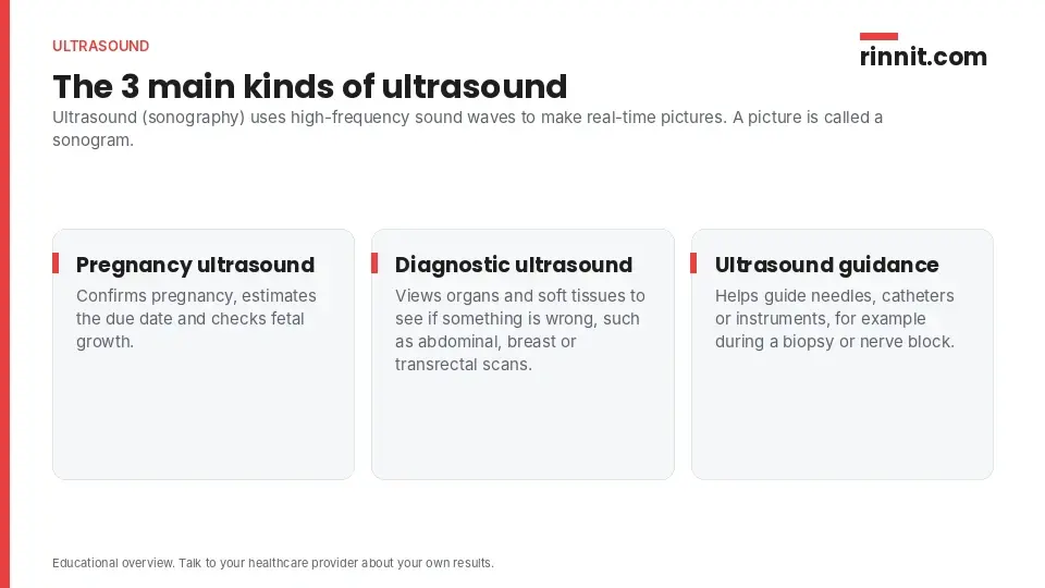
Type | What it is used for |
Pregnancy (prenatal) ultrasound | Confirming pregnancy, estimating the due date and checking fetal growth |
Diagnostic ultrasound | Viewing organs and tissues to find problems, such as abdominal, breast and transrectal ultrasounds |
Ultrasound guidance | Guiding needles, catheters or instruments during procedures such as a biopsy or nerve block |
Sources: [1]
2D, 3D and 4D ultrasound
Most ultrasound images are 2D, which produces a flat picture. A 3D ultrasound shows depth and angles that 2D cannot, giving more detailed images. A 4D ultrasound is a 3D ultrasound in motion, like a live 3D image. [1]
How does an ultrasound work?
A provider passes a device called a transducer (or probe) over your skin or inside a body opening. A thin layer of gel helps the sound waves travel from the probe into your body. [1]
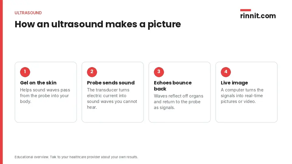
The probe turns electrical current into high-frequency sound waves that you cannot hear.
The waves travel into your tissues and bounce off structures inside your body.
The echoes return to the probe, which turns them into electrical signals.
A computer converts the signals into real-time images or video on a nearby screen.
Sources: [1]
How do you prepare for an ultrasound?
Preparation depends on the type of ultrasound. For example, you may need a full bladder for a pelvic ultrasound or an empty stomach for an ultrasound of your digestive tract. You may be asked to remove jewelry or clothing, or change into a gown. Some ultrasounds need no preparation at all, and your provider will tell you what to do. [1]
What happens during an ultrasound?
For a scan over the skin, the general steps are: [1]
You lie on your back or side on a comfortable table.
The technologist applies a small amount of water-soluble gel to the area. It does not harm your skin or stain your clothes.
The technologist glides a handheld probe over the gel to capture images.
You may be asked to stay very still or hold your breath for a few seconds for clearer pictures.
Once enough images are taken, the gel is wiped off and you are done.
If the probe goes inside your vagina or rectum, the steps are the same, except the probe is first covered with a sterile, lubricated cover so it slides in more easily. These internal scans may be uncomfortable but should not hurt. [1]
Is an ultrasound safe?
Yes. Research has largely shown ultrasound to be safe with no harmful side effects. Unlike X-rays and CT scans, it does not use radiation. It should still be performed by a professional trained to use it safely. [1]

Ultrasound | X-ray and CT scan | |
How it works | High-frequency sound waves | X-rays |
Radiation | None | Yes, ionizing radiation |
Sources: [1]
What can an ultrasound detect?
Ultrasound can help diagnose a wide range of problems, including: [1]
Abdominal aortic aneurysm
Abnormal growths, such as tumors or cancer
Blood clots
Gallbladder inflammation (cholecystitis) and gallstones
Ectopic pregnancy
Enlarged spleen
Kidney or bladder stones
Varicocele (enlarged veins in the scrotum)
What do ultrasound results mean?
You may get results quickly, or it may take several days for a radiologist to analyze the images. The radiologist sends a report to the provider who ordered the scan, and your provider shares the results with you. [1]
Report finding | What the wording usually means |
Normal | "No evidence" of a condition, an organ of "normal size," or conditions "not detected" |
Abnormal | Words like "evidence of" or "findings of," or the size of a mass or cyst |
Sources: [1]
If you are unsure what your report says, ask your provider to explain it. [1]
Questions to ask your provider
What type of ultrasound do I need?
What should I do to prepare?
Do I need any other tests?
When should I expect my results?
Conclusion
Ultrasound is one of the most common, safe and effective imaging tests. It can monitor a pregnancy, help find the cause of symptoms and guide procedures, all without radiation. Preparation is usually simple, and your provider will explain what your report means for you. [1]
Keep learning: Read our guides to kidney stones and ectopic pregnancy. For more on how ultrasound works, see the National Institute of Biomedical Imaging and Bioengineering's ultrasound overview.
FAQ
What is an ultrasound?
An ultrasound (sonography) is a noninvasive imaging test that uses high-frequency sound waves to create real-time pictures or video of organs and soft tissues [1].
What is an ultrasound used for?
It is used in pregnancy, to diagnose problems in organs and tissues, and to guide needles or instruments during procedures such as biopsies [1].
Is an ultrasound safe?
Yes. Research has largely shown ultrasound to be safe with no harmful side effects, and it does not use radiation [1].
Does an ultrasound hurt?
Ultrasounds over the skin are generally not painful. Scans inside a body opening may be uncomfortable but should not hurt [1].
How long does an ultrasound take?
Usually 30 minutes to an hour [1].
How do I prepare for an ultrasound?
It depends on the scan. You may need a full bladder for a pelvic ultrasound or an empty stomach for a digestive scan, and some scans need no preparation [1].
What can an ultrasound detect?
It can help diagnose gallstones, kidney and bladder stones, blood clots, tumors, an ectopic pregnancy, an enlarged spleen and an abdominal aortic aneurysm, among others [1].
How soon will I get ultrasound results?
Sometimes quickly, or it may take several days for a radiologist to review the images and report to your provider [1].
What is the difference between a 2D, 3D and 4D ultrasound?
2D shows flat images. 3D adds depth and angles for more detail, and 4D is 3D ultrasound in motion [1].
Who performs an ultrasound?
A doctor or a trained ultrasound technologist (sonographer) [1].
Related Reading
Testicular Ultrasound: Checking Lumps, Pain and Swelling explains the ultrasound used to check scrotal lumps, pain and swelling.
Learn more in our guide to Kidney Stones: Symptoms, Causes, and Treatment Guide.
Learn more in our guide to Ectopic Pregnancy: Symptoms, Causes, and Treatment.
Learn more in our guide to Deep Vein Thrombosis (DVT): Symptoms, Causes, Diagnosis & Treatment — Complete Guide.
Learn more in our guide to Choledocholithiasis: What a Bile Duct Gallstone Is, How It's Found, and How It's Removed.
Point-of-Care Ultrasound (POCUS): What It Is, Uses and How It Differs From a Standard Scan explains the bedside ultrasound clinicians use for quick, focused answers.
References
Cleveland Clinic. Ultrasound: Procedure Details & Results. Cleveland Clinic. Last updated March 5, 2026. https://my.clevelandclinic.org/health/diagnostics/4995-ultrasound
Yu SH, Kim CB, Park JW, et al. Ultrasonography in the diagnosis of appendicitis: evaluation by meta-analysis. Korean J Radiol. 2005;6(4):267-277. PMID 16374085. https://doi.org/10.3348/kjr.2005.6.4.267
Stengel D, Leisterer J, Ferrada P, et al. Point-of-care ultrasonography for diagnosing thoracoabdominal injuries in patients with blunt trauma. Cochrane Database Syst Rev. 2018;12(12):CD012669. PMID 30548249. https://doi.org/10.1002/14651858.CD012669.pub2
Medical disclaimer
This article is for general education only and does not replace advice from a qualified healthcare professional. Talk to your doctor about your own diagnosis and treatment, and seek emergency care right away for severe or sudden symptoms.



Comments