Retinal Imaging: Fundus Photos, OCT and Angiography Explained

Updated: 2 days ago
Written and medically reviewed by Dr. Baraa Alnahhal, MD · Last reviewed: November 2026 · How we review
Quick Answer
Retinal imaging is a group of tests that use special cameras and scanners to take detailed pictures of the back of the eye, including the retina, macula and optic nerve. It helps detect and track diabetic retinopathy, macular degeneration, glaucoma and retinal detachment. You'll usually get dilating drops, so plan for a ride home.
What the research says
Wide-field photos can work without dilation: in people with diabetic retinopathy, gradings from a nondilated 200-degree ultra-widefield camera agreed well with standard dilated seven-field photography (kappa about 0.66–0.74) while covering a larger area of the retina (Kernt M et al., Ophthalmologe, 2011) [2]
Serious dye reactions are rare: a review of 78 studies on fluorescein angiography found severe reactions in about 0.04%–0.59% of people, and that a past reaction to fluorescein was the only proven predictor of another reaction (Kornblau IS, El-Annan JF, Surv Ophthalmol, 2019) [3]
Key takeaways
Detailed images of the retina, macula and optic nerve.
Main types: fundus photography, OCT and fluorescein angiography.
Finds diabetic eye disease, macular degeneration, glaucoma and more.
Dilating drops are usually used, so arrange a ride home.
Repeat images help track changes over time.
What is retinal imaging?
Retinal imaging captures the fundus, the inside back surface of the eye. It's used during comprehensive eye exams and follow-up visits. Repeat images over time help your eye doctor spot subtle changes and see how well treatment is working. Learn more about the retina. Sources: [1]
Types of retinal imaging

Each type shows the eye in a different way.
Type | What it shows | Time |
Color photos of the retina and blood vessels, sometimes wide-field | About 5–10 minutes | |
Cross-section scans and thickness of retina layers | About 5–10 minutes | |
Dye in an arm vein highlights leaks and blockages in eye vessels | Up to 30 minutes |
Sources: [1]
What can it find?
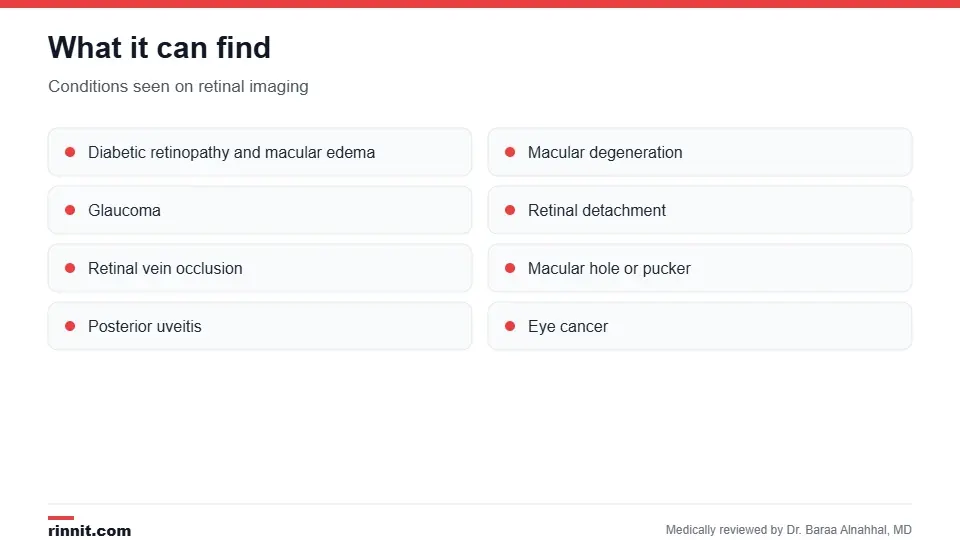
Retinal imaging helps detect many eye diseases.
Diabetic retinopathy and diabetic macular edema.
Macular degeneration, macular hole and macular pucker.
Retinal vein occlusion and posterior uveitis.
Eye cancer. Sources: [1]
How do I prepare?
Your provider may give you dilating drops, since a wider pupil gives better images. Arrange a ride home, because your vision may be blurry and sensitive to light for a few hours. Sources: [1]
What happens during the test?
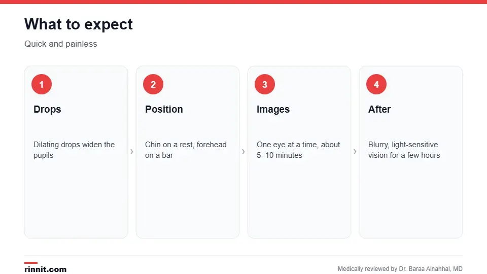
Retinal imaging is quick and painless.
You sit at the device with your chin on a chin rest and forehead against a bar.
Images are taken one eye at a time; you may be asked to look at a green light, which can feel briefly bright.
For fluorescein angiography, dye is injected into a vein in your arm and photos are taken as it moves through the eye's vessels. Nothing touches your eye. Sources: [1]
Afterward, avoid driving, reading and screens for a few hours if your eyes were dilated, and wear sunglasses outdoors. Sources: [1]
Risks
Fundus photography and OCT have no known risks because they use only a camera. Fluorescein angiography is low risk; your skin may look yellow and your urine dark yellow or orange for up to 24 hours. Rarely, it causes nausea, itching or hives, and severe allergic reactions are extremely rare. Sources: [1, 3]
Understanding your results
Your eye doctor reviews the images, often with you. They become part of your record so new images can be compared with old ones to track changes. Sources: [1, 2]
Conclusion
Retinal imaging gives your eye doctor a clear, lasting view of the back of your eye. It's one of the best tools for catching diabetic eye disease, macular degeneration and glaucoma early and for tracking treatment.
Keep learning: read about retinal disease and OCT scans.
Frequently asked questions
What is retinal imaging?
Detailed pictures or scans of the back of the eye.
What are the main types?
Fundus photography, OCT and fluorescein angiography.
Does it hurt?
No. Most types use only a camera.
Will my eyes be dilated?
Usually, yes.
Can I drive afterward?
Not if your eyes were dilated; arrange a ride.
How long does it take?
About 5–10 minutes, or up to 30 minutes for angiography.
What can it detect?
Diabetic retinopathy, macular degeneration, glaucoma and more.
Is fluorescein dye safe?
Yes, for most people; serious reactions are rare.
Why does my urine turn orange?
The dye leaves your body through urine within about a day.
Why repeat the images?
To compare and track changes over time.
Related Reading
Amsler Grid Eye Test: How to Check Your Central Vision at Home explains the at-home grid used to check central vision.
Fundus Photography: What It Shows, What to Expect and Who Needs It color photos of the back of the eye.
Optical Coherence Tomography (OCT) Eye Test: What It Shows and What to Expect cross-section scans of the retina.
Diabetic Retinopathy: Symptoms, Causes, Diagnosis & Treatment — Complete Guide a common reason for retinal imaging.
Eye Exam: What Happens, Tests Included and How Often to Go where retinal imaging often happens.
What Is Retinal Disease? A Plain-Language Guide to the Back of Your Eye, Its Warning Signs, and Its Treatment conditions that affect the retina.
References
Cleveland Clinic. Retinal Imaging: Purpose & Procedure Details. Cleveland Clinic. Last updated June 5, 2023. https://my.clevelandclinic.org/health/diagnostics/25046-retinal-imaging
Kernt M, Pinter F, Hadi I, et al. Diabetic retinopathy: comparison of the diagnostic features of ultra-widefield scanning laser ophthalmoscopy Optomap with ETDRS 7-field fundus photography. Ophthalmologe. 2011;108(2):117-123. PMID 20683601. https://doi.org/10.1007/s00347-010-2226-4
Kornblau IS, El-Annan JF. Adverse reactions to fluorescein angiography: a comprehensive review of the literature. Surv Ophthalmol. 2019;64(5):679-693. PMID 30772364. https://doi.org/10.1016/j.survophthal.2019.02.004
Medical disclaimer
This article is for general education only and does not replace advice from a qualified healthcare professional. Talk to your doctor about your own diagnosis and treatment, and seek emergency care right away for severe or sudden symptoms.



Comments