Retina: Function, Anatomy, Symptoms, Tests and Care

Updated: 3 days ago
Medically reviewed by Dr. Baraa Alnahhal, MD · Last reviewed: October 2026
Quick Answer: What does the retina do?
The retina senses light and converts it into coded nerve signals. Those signals travel along the optic nerve to the brain, where they are processed as vision. It sits at the back of the eyeball, behind the iris and lens, and light rays converge on it when the eye focuses properly. [1]
Question | Short answer |
Where is the retina? | At the back of the eyeball, behind the iris and lens. |
What are its main regions? | The macula supports color and fine-detail vision. The peripheral retina supports side vision, and its rods help with low-light vision. |
When is it urgent? | Sudden vision loss or sudden new vision changes that begin over minutes or a few hours need emergency care. |
What the research says
Diabetic retinopathy affects roughly 1 in 5 people with diabetes. A systematic review and meta-analysis of 59 population-based studies found a global prevalence among people with diabetes of 22.27% (95% CI 19.73–25.03%) for diabetic retinopathy, 6.17% (95% CI 5.43–6.98%) for vision-threatening diabetic retinopathy and 4.07% (95% CI 3.42–4.82%) for clinically significant macular edema. The number of adults with diabetic retinopathy was estimated at 103.12 million in 2020, projected to reach 160.50 million by 2045 (Teo et al., Ophthalmology, 2021). [2]
Age-related macular degeneration is common worldwide and expected to rise. A systematic review and meta-analysis of 39 studies including 129,664 individuals found a pooled prevalence of any age-related macular degeneration of 8.69% (95% credible interval 4.26–17.40%) when mapped to ages 45–85. The number of people affected was projected to rise from 196 million in 2020 to 288 million in 2040 (Wong et al., Lancet Glob Health, 2014). [3]

What is the retina?
The retina is a layer of cells at the back of the eyeball. It acts as the eye's light-sensitive interface: it converts incoming light into nerve signals and sends those signals through the optic nerve to the brain. The brain then processes them into your sense of vision. [1]
This conversion is possible because the retina contains light-detecting cells called photoreceptors, which convert light into coded signals that the brain can decode. A helpful way to think about the pathway is light → retina → optic nerve → brain → vision. [1]
How does the retina support vision?
The retina does not produce the finished image by itself. It senses light and sends coded information onward, and the brain receives, decodes and organizes those signals into the big-picture view you experience.
Because this pathway is central to sight, retinal damage can change the way the world looks, leave gaps in vision or cause total blindness. Many conditions that affect the retina can cause permanent damage and vision loss when they are not treated quickly.
Part of the visual pathway | Role |
Retina | Converts light into nerve signals. |
Photoreceptors | Convert light into coded signals. |
Optic nerve | Carries retinal signals to the brain. |
Brain | Decodes the signals into vision. |
What are the macula and peripheral retina in retina anatomy?
The retina has two main parts: the macula and the peripheral retina. They contribute to different parts of the visual experience. [1]
Retinal region | Main visual contribution |
Macula | Cones are essential for color and fine-detail vision, which supports reading, seeing faces and driving. |
Peripheral retina | Allows side vision when looking straight ahead. Rods in this region also help with low-light vision. |

What conditions can affect the retina?
Many conditions that damage the eye can affect the retina. Retinal diseases and disorders include conditions involving the macula, blood vessels, inflammation, the vitreous, tumors and inherited or developmental changes, including retinal detachment.
Group | Examples |
Macular conditions | Age-related macular degeneration, macular hole and macular pucker. |
Blood-vessel or systemic-related conditions | Diabetes-related retinopathy, hypertensive retinopathy, retinal bleeding, retinal vein occlusion and retinal artery occlusion (eye stroke). |
Retinal separation, inflammation or vitreous-related conditions | Retinal tears, retinal detachments, retinal inflammation (uveitis) and posterior vitreous detachment. |
Inherited, developmental or light-related conditions | Retinitis pigmentosa, retinopathy of prematurity, solar retinopathy, color blindness and achromatopsia. |
Tumors | Eye cancers such as retinoblastoma and benign tumors. |
Other | Ocular migraine. |
This list is not a way to identify a condition from a symptom. Similar visual changes can occur in different situations, and a professional eye examination is needed to interpret them.
What are the possible symptoms of a retina problem?
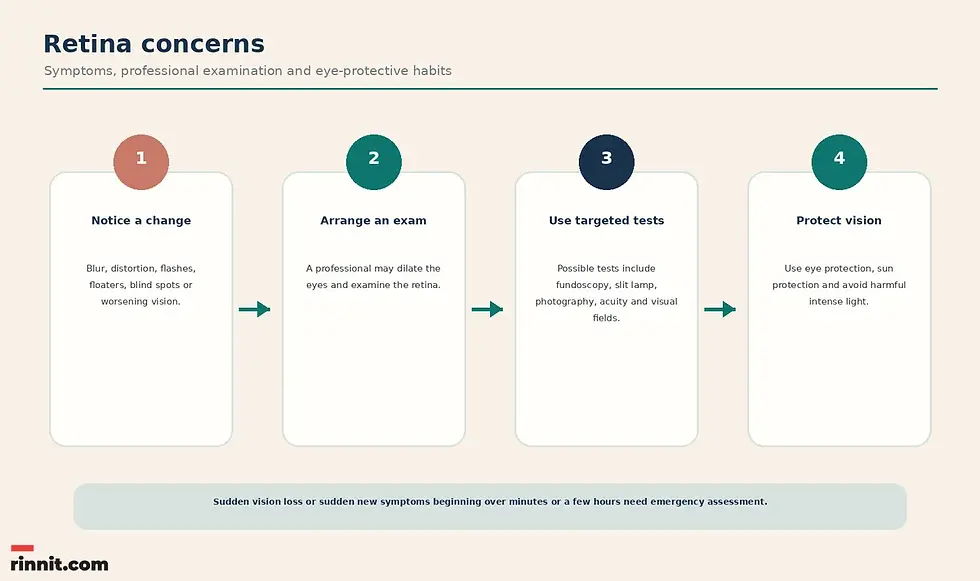
Talk with a healthcare professional if you notice any visual change. Possible symptoms include blurred or distorted vision, peripheral vision loss or tunnel vision, double vision, flashes, floaters, light sensitivity, blind spots or visual field defects, and vision that is becoming noticeably worse.
Symptom | Plain-language description |
Blurry or distorted vision | Details look unfocused, warped or changed. |
Peripheral vision loss | Side vision narrows or becomes reduced. |
Double vision | One object appears as two. |
Flashes | Brief flashes or streaks of light appear in the visual field. |
Floaters | Spots, strands or shapes move across the visual field. |
Light sensitivity | Light feels unusually uncomfortable. |
Blind spots or field defects | A missing or blocked area appears in the visual field. |
Noticeably worsening vision | Vision becomes progressively or clearly less effective. |
A symptom does not establish a diagnosis. Timing matters: sudden vision loss or a sudden new visual change that develops over minutes or a few hours should be treated as an emergency. [1]
How do professionals examine the retina?
A healthcare professional checks the retina as part of an overall eye exam, and the eyes are dilated for parts of it. Retina-specific parts of an eye exam include the tests below. [1]
Test or examination | What it is |
Fundoscopy or ophthalmoscopy | Direct examination of the retina. |
Slit-lamp examination | A retina-related part of the eye examination. |
Fundus photography | Photography of the back of the eye. |
Fundus reflex (red reflex) testing | Testing of the fundus reflex. |
Visual acuity testing | Assessment of how clearly you see. |
Visual field testing | Assessment of the field of vision. |
The appropriate combination of examinations depends on the professional's question, your symptoms and your overall eye health. Results should be interpreted by the professional who ordered or performed the examination.
How can you support retinal and eye health?
Everyone needs regular eye examinations, whether or not they wear glasses or contact lenses, at least every one to two years. People with chronic conditions that can affect the eyes, such as diabetes, should see an eye care provider at least once a year. An eye care specialist may recommend a more frequent schedule when needed.
Protective habit | Why it helps |
Keep regular eye appointments | Retinal changes can be identified as part of eye care. |
Prioritize nutrition and a healthy-for-you weight | Listed as eye-health precautions. |
Use protective eyewear during risky activities | Eye protection can reduce injury risk. |
Use sun protection | Sunglasses labeled as blocking 99% to 100% of UVA and UVB rays help protect the eyes; polarization reduces glare but does not itself block UV. |
Avoid dangerous intense light | Light sources such as welding flames and burning magnesium can permanently harm the retina. |
Do not ignore vision changes | Even gradual changes deserve professional review. |
When should you seek care?
See a healthcare professional as soon as you notice changes in your vision. Do not wait for symptoms to get worse, whether the change turns out to be something as simple as needing new glasses or something more serious.
Go to an emergency room for sudden vision loss or the sudden appearance of new vision changes or symptoms that start or develop over minutes or a few hours. A sudden increase in floaters, new flashes of light, or a shadow or curtain over part of your vision can be signs of a retinal tear or detachment and need same-day eye care. These changes can occur with conditions that quickly cause irreversible damage, partial vision loss or blindness. [1]
Conclusion
The retina is the light-sensitive layer at the back of the eye that converts light into signals for the brain. Its macula supports color and fine-detail vision, while the peripheral retina supports side vision and low-light vision. Because retinal problems can affect sight permanently, new visual changes deserve professional attention, and sudden changes require emergency assessment. [1]
If your vision has changed, arrange an eye examination rather than trying to identify the cause from symptoms alone. Bring a clear description of when the change started, whether it affects one or both eyes, and whether it involves flashes, floaters, distortion, blind spots or worsening clarity. [1]
Frequently Asked Questions
What is the retina?
The retina is a layer of cells at the back of the eyeball. It converts light into nerve signals and sends those signals through the optic nerve to the brain, where they are processed into vision.
Is the retina behind the lens?
Yes. The retina is at the back of the eye, behind the iris and lens. When the eye focuses properly, light rays converge on the retina.
What are the two main parts of the retina?
The macula and the peripheral retina.
What is the macula responsible for?
The macula contains cones that are essential for color and fine-detail vision. This type of vision supports activities such as reading, seeing faces and driving.
What does the peripheral retina do?
The peripheral retina allows you to see to the side when looking straight ahead. Rods in the peripheral retina also help with low-light vision.
What are photoreceptors?
Photoreceptors are light-detecting cells in the retina that convert light into coded signals the brain can decode.
What conditions can affect the retina?
Examples include age-related macular degeneration, diabetes-related retinopathy, hypertensive retinopathy, macular hole, macular pucker, retinal tears and detachments, retinal vein or artery occlusion, uveitis, retinitis pigmentosa, retinopathy of prematurity, solar retinopathy, eye cancers and color blindness.
What are possible symptoms of a retinal problem?
Symptoms can include blurry or distorted vision, peripheral vision loss, double vision, flashes, floaters, light sensitivity, blind spots or visual field defects, and noticeably worsening vision.
Are flashes and floaters always a retinal emergency?
A symptom alone does not identify its cause. Sudden new vision changes or sudden vision loss that develop over minutes or a few hours need emergency assessment.
What tests can examine the retina?
An eye specialist may use dilation, fundoscopy or ophthalmoscopy, a slit-lamp exam, fundus photography, red-reflex testing, visual acuity testing and visual field tests as part of an eye exam.
How often should I have an eye exam?
At least every one to two years for everyone, and at least once a year for people with chronic conditions that can affect the eyes, such as diabetes. An eye care specialist may recommend a different schedule.
Can I prevent retinal problems?
You can support eye health with regular eye exams, a weight that is healthy for you, proper nutrition, eye protection during risky activities, sun protection and avoiding light sources bright enough to cause permanent harm. Do not ignore vision changes, even gradual ones.
When should I go to the emergency room?
Go to the emergency room for sudden vision loss or the sudden appearance of new vision changes or symptoms that start or develop over minutes or a few hours.
Related Reading
Retinal Detachment: Symptoms, Causes, Diagnosis & Treatment — Complete Guide explains the warning signs of a detached retina and urgent repair.
Diabetic Retinopathy: Symptoms, Causes, Diagnosis & Treatment — Complete Guide covers how diabetes damages retinal blood vessels and how to protect vision.
The Macula: The Center of Detailed Vision describes the central retina that provides sharp, detailed vision.
Optical Coherence Tomography (OCT) Eye Test: What It Shows and What to Expect explains the eye scan that shows the retina's layers in detail.
References
Cleveland Clinic. Retina of the Eye: What It Is, Function & Anatomy. Cleveland Clinic. Updated October 3, 2024. https://my.clevelandclinic.org/health/body/22694-retina-eye
Teo ZL, Tham YC, Yu M, et al. Global Prevalence of Diabetic Retinopathy and Projection of Burden through 2045: Systematic Review and Meta-analysis. Ophthalmology. 2021;128(11):1580-1591. PMID 33940045. https://doi.org/10.1016/j.ophtha.2021.04.027
Wong WL, Su X, Li X, et al. Global prevalence of age-related macular degeneration and disease burden projection for 2020 and 2040: a systematic review and meta-analysis. Lancet Glob Health. 2014;2(2):e106-e116. PMID 25104651. https://doi.org/10.1016/S2214-109X(13)70145-1
Medical disclaimer
This article is for general educational purposes and is not a substitute for professional medical advice, diagnosis or treatment. Always seek the advice of a qualified healthcare professional with any questions about a medical condition or treatment.



Comments