Photoreceptors: Rods, Cones, Vision and Eye Health

Updated: 3 days ago
Medically reviewed by Dr. Baraa Alnahhal, MD · Last reviewed: October 2026
Quick Answer: what are photoreceptors?
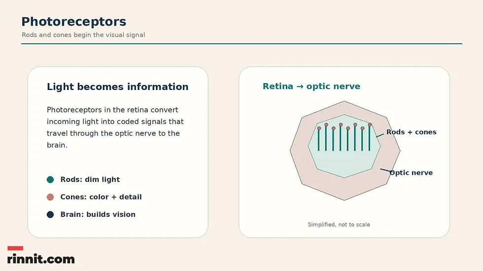
Photoreceptors are light-sensitive neurons in the retina. Rods respond especially well to very low light, while cones require more light and support color and detailed central vision. Together, they begin the process that turns light entering the eye into coded nerve signals the brain uses for sight. [1]
Core idea | Plain-language meaning |
Location | On the retina at the back of the eye. |
What they detect | Incoming light. |
What they produce | Coded nerve signals sent through the optic nerve. |
Rods | Low-light and night vision; no color detection. |
Cones | Color vision and fine detail in brighter light. |
Main clinical symptom | Vision loss that can take different forms. |
What the research says
Retinitis pigmentosa shows how rods and cones can fail in sequence. A comprehensive review describes retinitis pigmentosa as a group of inherited retinal dystrophies marked by primary degeneration of rod and cone photoreceptors, with a worldwide prevalence of about 1 in 4,000. It typically begins with night blindness in adolescence, followed by concentric visual field loss (reflecting rod dysfunction), with central vision loss later in life due to cone dysfunction. Electroretinogram responses are markedly reduced or absent, and variants in more than 80 genes have been linked to the non-syndromic form (Verbakel et al., Prog Retin Eye Res, 2018). [2]
Color vision deficiency is more common in males than in females. A systematic review and meta-analysis of 56 studies (1,703,619 participants) estimated a global prevalence of congenital color vision deficiency of 2.59% in children and adolescents: 4.38% in males versus 0.64% in females. Deutan defects were the most common type (Jeong et al., Ophthalmology, 2025). [3]
Where are photoreceptors located?
Photoreceptors are on the retina, a light-sensitive layer at the back of the eye. The retina and brain work together: retinal photoreceptors start the process, the optic nerve carries coded signals and the brain decodes those signals to build what a person sees. [1]
Because of how the retina develops, photoreceptors are technically part of the central nervous system, along with the brain and spinal cord. They are a specialized type of neuron that uses electrical and chemical signaling. [1]
How do photoreceptors create vision?
When light enters the eye and reaches rods and cones, chemical and electrical processes activate within the receptors and connected retinal cells. These processes convert light into signals the brain can use. The retina sends coded nerve signals through the optic nerve to the brain, which decodes them and uses them to build the visual experience. [1]
A digital-camera comparison can help explain the sequence: a camera sensor detects light and converts it into computer code, while the retina detects light and converts it into nerve signals. The comparison is only an analogy; human vision involves living retinal and nervous-system tissues. [1]
Stage | What happens |
1. Light enters | Light reaches the retina at the back of the eye. |
2. Photoreceptors respond | Rods and cones activate chemical and electrical processes. |
3. Signals are coded | Retinal cells convert detected light into nerve signals. |
4. The optic nerve carries information | Signals travel toward the brain. |
5. The brain decodes | The brain uses the signals to build what you see. |
What are rods?
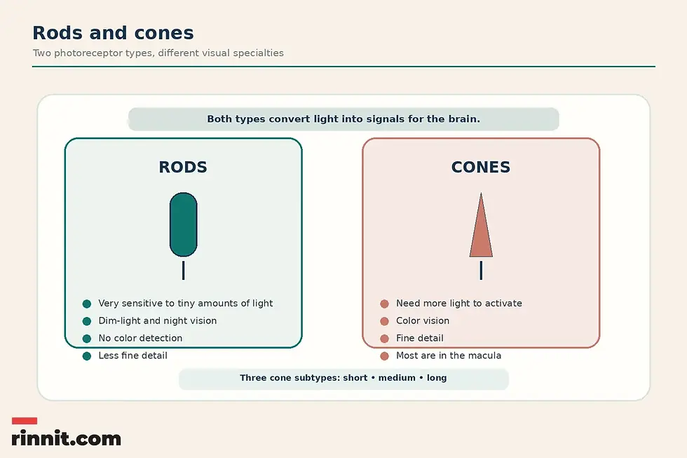
Rods are tall, cylindrical or tubelike photoreceptors. They are extremely sensitive to tiny amounts of light, which makes them especially useful in dim settings. [1]
About 95% of photoreceptors are rods, or about 100–125 million cells. Rods help with low-light and night vision but are not as good at fine detail and cannot detect colors. [1]
Rod-based vision in dim light is called scotopic vision. Conditions affecting rods often involve some form of night blindness, also called nyctalopia, early in their course. [1]
What are cones?
Cones are photoreceptors with a cone-like shape: circular at the bottom and pointed at the top. They need more light than rods to activate, but they can detect colors when active. [1]
Cones are packed most densely in the macula, especially its center, the fovea. This concentration helps explain why the center of the visual field is used for color and fine detail.
Conditions involving cones may cause color blindness or gradual loss of color vision. They may also reduce fine-detail vision, making images appear cloudy or fuzzy. [1]
Rods versus cones: what is the difference?
Rods and cones are both retinal photoreceptors, but they specialize in different lighting and visual tasks.
Feature | Rods | Cones |
Shape | Tall and cylindrical. | Cone-like, with a pointed tip. |
Light sensitivity | Extremely sensitive to tiny amounts of light. | Need more light to activate. |
Main role | Dim-light and night vision. | Color vision and fine detail. |
Color detection | Cannot see colors. | Detect color-related wavelengths. |
Number and location | About 95% of photoreceptors (roughly 100–125 million). | Most densely packed in the macula. |
Associated changes | Night blindness may appear early in rod-related conditions. | Color-vision loss and cloudy or fuzzy detail may occur in cone-related conditions. |
How does color vision work?
The eyes have three main cone types that work together for color vision. The cones do not see colors by themselves. Instead, they respond to light wavelengths and send information to the brain. The brain compares the signals from the three cone types and turns them into color perception. [1]
The three cone subtypes are short, medium and long, sometimes described as blue, green and red. Their sensitivity overlaps, so color perception depends on comparing their combined signals rather than using one cone type in isolation. [1]
Having three cone subtypes is called trichromacy. The average healthy human eye can distinguish up to 1 million colors. [1]
Cone subtype | Common description |
Short | Blue. |
Medium | Green. |
Long | Red. |
What is tetrachromacy?
Tetrachromacy involves four cone subtypes rather than three. It arises from a rare genetic mutation that can only happen in females. [1]
In weak tetrachromacy, the brain cannot fully process the fourth cone's input. In strong tetrachromacy, the brain does process it. The fourth cone may enable distinction of 100 million colors, but strong tetrachromacy is very rare, making its frequency and further research difficult to establish. [1]
What conditions affect rods and cones?
Rods and cones can be affected by several eye conditions, including color blindness, cone-rod dystrophy, macular degeneration and retinitis pigmentosa. Some also affect other retinal structures or surrounding eye tissue. Named conditions include partial or total color blindness (including achromatopsia), cone-rod dystrophy, macular degeneration, Refsum disease, retinitis pigmentosa, solar retinopathy and Usher syndrome. [1]
Condition or group | Association with photoreceptors |
Color blindness | May be partial or total, with achromatopsia as the total form. |
Cone-rod dystrophy | A photoreceptor-related condition. |
Macular degeneration | Can affect rods and cones, especially when retinal tissue is damaged or displaced. |
Refsum disease | A photoreceptor-related condition. |
Retinitis pigmentosa | Rod-related symptoms may occur early and later involve cones. |
Solar retinopathy | A photoreceptor-related condition. |
Usher syndrome | A photoreceptor-related condition. |
What symptoms can photoreceptor conditions cause?
Vision loss is the main symptom of a photoreceptor-related condition, but the pattern depends on which cells are affected.
Rod-related conditions usually cause some form of night blindness early on. Some conditions, including retinitis pigmentosa, can worsen and later affect cone photoreceptors too. [1]
Cone-related conditions often involve color blindness or loss of color vision over time. They may also affect fine detail, causing vision to seem cloudy or fuzzy. Some can progress and later cause rod-related symptoms. [1]
Many conditions affect both rods and cones, particularly when retinal tissue is damaged or displaced. Macular degeneration is a key example. These patterns can provide questions to discuss with an eye-care professional, but they cannot diagnose a condition by themselves.
How are rods and cones tested?
A standard eye examination allows an eye-care specialist to inspect the back of the eye for retinal changes or other tissue changes. Parts of the examination, especially dilation and the slit-lamp examination, can identify conditions that may otherwise be invisible and may not cause early symptoms. [1]
More specific testing may include electroretinography, which measures electrical activity in the retina, and visual evoked potentials (VEP). VEP is a brain test that can show whether signals from the retina are reaching the brain. [1]
An eye-care specialist may also recommend imaging to view retinal layers or map blood vessels in and around the retina. This may help identify new blood-vessel growth or tissue changes that could lead to retinal damage or vision loss. [1]

Test or examination | What it may show |
Standard eye exam | Changes at the back of the eye and other retinal tissue changes. |
Eye dilation | Conditions otherwise invisible or without early symptoms. |
Slit-lamp exam | Conditions that may be difficult to detect without magnification. |
Electroretinography | Electrical activity in the retina. |
Visual evoked potentials | Whether retinal signals reach the brain. |
Retinal imaging | Retinal layers, blood vessels, new vessel growth or tissue changes. |
How can you support retinal and eye health?
Everyone, including people who do not need glasses or contact lenses, should have an eye exam at least every two years, or more often when a condition requires closer monitoring. [1]
Regular physical examinations can help identify chronic conditions that may affect the eyes, especially high blood pressure and type 2 diabetes, before symptoms or eye damage begin. [1]
Avoid nicotine, including smoking, vaping and smokeless tobacco. Blood vessels supplying the retina are particularly vulnerable to changes associated with nicotine use. [1]
Nutrition matters for overall health, and vitamin A plays an important role in how photoreceptors detect light. Reaching and maintaining a weight that is healthy for you also helps. [1]
Eye-health topic | Guidance |
Eye examinations | At least every two years for everyone, or more often when closer monitoring is needed. |
Physical examinations | Help identify conditions such as high blood pressure and type 2 diabetes. |
Nicotine | Avoid smoking, vaping and smokeless tobacco. |
Nutrition | Prioritize nutrition; vitamin A supports photoreceptor light detection. |
Weight | Reach and maintain a weight that is healthy for you. |
When should you contact an eye-care professional?
Discuss new or changing vision, night-vision difficulty, loss of color vision, cloudy or fuzzy detail, or other persistent visual concerns with an eye-care professional. A person can have retinal conditions without early symptoms, which makes routine examinations useful.
Get emergency care right away for sudden vision loss, a curtain or shadow over part of your vision, or a sudden shower of new floaters or flashes of light, because these can signal a retinal detachment, stroke or other emergency. This article cannot determine the cause or urgency of an individual symptom.
Conclusion: rods and cones begin the visual signal
Photoreceptors are specialized neurons in the retina that turn light into coded signals for the brain. Rods are highly sensitive in dim light and support night vision, while cones work in brighter light to support color and fine detail.
The brain compares signals from three cone subtypes to create color perception, and the optic nerve carries retinal information toward the brain. Conditions that affect rods, cones or surrounding retinal tissue can cause different forms of vision loss. Regular eye examinations, attention to overall health and professional evaluation of changing vision can support informed eye care.
Frequently Asked Questions
What are photoreceptors?
Photoreceptors are specialized light-detecting neurons on the retina at the back of the eye. They convert light into signals the brain can use for vision.
What are rods?
Rods are cylindrical retinal photoreceptors that are highly sensitive to small amounts of light. They support dim-light and night vision but cannot detect colors.
What are cones?
Cones are cone-shaped photoreceptors that need more light than rods. They detect color-related wavelengths and support fine detail, especially in the center of the visual field.
What is the difference between rods and cones?
Rods specialize in low-light vision and do not detect color. Cones need more light but support color vision and fine detail.
Where are cones concentrated?
Cones are most densely packed in the macula, a part of the retina associated with central color and fine-detail vision.
How many rods are in the human eye?
About 100–125 million, making up about 95% of photoreceptors.
What is scotopic vision?
Scotopic vision is vision in dim light that relies mainly on rod photoreceptors.
How does the eye see color?
Three cone subtypes respond to overlapping ranges of light wavelengths. The brain compares their signals and turns them into color perception.
What is trichromacy?
Trichromacy is having three cone subtypes: short, medium and long.
What is tetrachromacy?
Tetrachromacy involves four cone subtypes. It comes in weak and strong forms, and strong tetrachromacy is very rare.
What conditions affect photoreceptors?
Named conditions include color blindness, achromatopsia, cone-rod dystrophy, macular degeneration, Refsum disease, retinitis pigmentosa, solar retinopathy and Usher syndrome.
What is the main symptom of a photoreceptor condition?
Vision loss, though the form can vary. Rod-related conditions may cause night blindness, while cone-related conditions may affect color and fine detail.
What is nyctalopia?
Nyctalopia is night blindness, which often appears early in rod-related conditions.
Can photoreceptor conditions affect both rods and cones?
Yes. Many conditions affect both types, especially when retinal tissue is damaged or displaced.
What tests check rods and cones?
An eye exam, dilation, slit-lamp examination, electroretinography, visual evoked potentials and retinal imaging may help evaluate the retina and visual signaling.
How often should I have an eye exam?
At least every two years, or more often when closer monitoring is needed.
Why does vitamin A matter for photoreceptors?
Vitamin A plays a vital role in how photoreceptors detect light.
When should I seek care for vision changes?
Discuss new, persistent or changing visual symptoms with an eye-care professional. Sudden or rapidly worsening vision changes need prompt professional assessment.
Related reading
What is retinal disease? A plain-language guide to the back of your eye
Color blindness (color vision deficiency): symptoms, causes, diagnosis and living tips
Dry macular degeneration: complete guide to symptoms, causes and treatment
Understanding retinitis pigmentosa (RP): causes, symptoms and management
Choroideremia: the inherited eye condition that starts with night blindness
References
Cleveland Clinic. Photoreceptors (Rods & Cones): Anatomy & Function. Cleveland Clinic. Updated May 6, 2024. https://my.clevelandclinic.org/health/body/photoreceptors-rods-and-cones
Verbakel SK, van Huet RAC, Boon CJF, et al. Non-syndromic retinitis pigmentosa. Prog Retin Eye Res. 2018;66:157-186. PMID 29597005. https://doi.org/10.1016/j.preteyeres.2018.03.005
Jeong YD, Cho J, Son Y, et al. Global Prevalence of Congenital Color Vision Deficiency among Children and Adolescents, 1932-2022. Ophthalmology. 2025;132(12):1431-1444. PMID 40769301. https://doi.org/10.1016/j.ophtha.2025.07.031
Disclaimer
This article is for general educational purposes and is not a substitute for professional medical advice, diagnosis or treatment. Always seek the advice of a qualified healthcare professional with any questions about a medical condition or treatment.



Comments