The Macula: The Center of Detailed Vision

Updated: 3 days ago
Medically reviewed by Dr. Baraa Alnahhal, MD · Last reviewed: October 17, 2026
Quick Answer
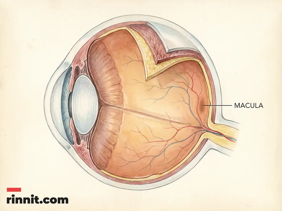
The macula is a round, yellow spot at the center of the retina at the back of your eye [1]. It processes central vision, letting you see objects directly in front of you in sharp detail [1]. It supports reading, driving, and recognizing colors or faces. By converting light into electrical signals for the brain [1], it lets you see fine detail.
What the research says
Antioxidant vitamins and minerals can slow progression to late AMD, but lutein and zeaxanthin alone show little effect. A Cochrane review of 19 trials found that people taking antioxidant vitamins were less likely to progress to late age-related macular degeneration (odds ratio 0.72, 95% CI 0.58–0.90; 2,445 participants), while lutein or zeaxanthin supplements gave a similar or only slightly reduced risk (risk ratio 0.94, 95% CI 0.87–1.01; 6,891 eyes, low-certainty evidence). (Evans & Lawrenson, Cochrane Database Syst Rev, 2017) [2]
Age, smoking and drusen predict who progresses. A meta-analysis of 18 cohort studies (38,697 people) found higher odds of AMD progression with each 5 years of age (odds ratio 1.14, 95% CI 1.09–1.19), current smoking (odds ratio 1.28, 95% CI 1.07–1.52) and extensive drusen covering more than 10% of the grid (odds ratio 7.85, 95% CI 4.66–13.23). (Tian & Zhang, Front Med, 2025) [3]
Smoking is strongly linked to developing AMD. A meta-meta-analysis of 12 qualifying reviews reported that current smokers had markedly higher odds of AMD than non-smokers (odds ratio 11.93, 95% CI 4.40–32.33; risk ratio 7.45, 95% CI 4.09–13.57) and past smokers a seven-fold increase (odds ratio 7.09, 95% CI 4.79–10.51). (Asiamah et al., Eur J Ophthalmol, 2025) [4]
What does the macula do for detailed vision?
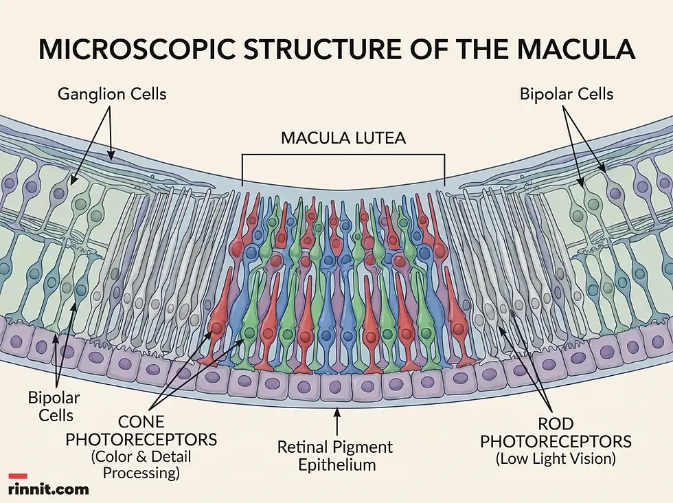
The macula acts as the eye’s primary processor for detailed imagery. While the rest of the retina handles peripheral vision and motion, the macula focuses on what is directly in front of the viewer. It translates light into images by utilizing specialized photoreceptor cells called cones and rods [1]. Cones are particularly concentrated in the macula [1], enabling the perception of vivid colors and fine details, such as the text on this page or the subtle features of a person’s face.
This tiny area works like a high-definition camera lens. When light enters the eye, it passes through the lens and hits the macula, where photoreceptors convert light energy into electrical signals [1]. These signals travel through the optic nerve to the brain [1], which then interprets them as a clear, focused picture. Without a healthy macula, vision would be blurry and lack the precision required for most daily tasks [1], even if the rest of the eye remains functional.
Function | Description | Daily Impact |
|---|---|---|
Central Vision | Processes objects directly in front of the eye. | Driving, walking, and navigation. |
Detail Processing | Captures fine lines and small movements. | Reading, sewing, and intricate work. |
Color Perception | High concentration of cones identifies hues. | Distinguishing between colors and shades. |
Facial Recognition | Focuses on specific features and expressions. | Social interaction and security. |
What is the anatomy and composition of the macula?
The macula, also known as the macula lutea [1], is a small, yellow, oval-shaped spot measuring approximately 5 millimeters in width [1]. Despite its size—less than a quarter of an inch [1]—it contains a high concentration of cone cells [1]. These cones are responsible for color vision and high-resolution detail. Rods are also present, primarily helping with vision in dim light and at night [1], but the macula’s primary strength lies in its ability to handle intense detail under bright conditions.
The yellow color of the macula comes from pigments that act as a natural “sunblock,” protecting the sensitive photoreceptors from high-energy blue and ultraviolet light. This anatomical design is crucial for maintaining the longevity of the cells. Because the macula is located at the very center of the retina, it is the direct target for light entering through the pupil, making its structural integrity paramount for maintaining a clear line of sight.
Anatomical Feature | Detail | Purpose |
|---|---|---|
Size | ~5 millimeters [1] | Compact area for focused processing. |
Color | Yellow (Macula Lutea) [1] | Pigments protect against UV/blue light. |
Cell Type | High concentration of cones | Enables high-definition and color vision. |
Location | Center of the retina [1] | Directly processes central light rays. |
What conditions affect the macula, and how can you care for it?
Conditions that affect the macula, such as age-related macular degeneration, can cause central vision loss, while protective habits and regular eye exams support macular health. The most well-known is age-related macular degeneration (AMD), which involves the gradual breakdown of the macula’s light-sensitive cells. Other issues include macular holes, puckers (epiretinal membranes), and cystoid macular edema. Because these conditions often develop gradually, noticing early symptoms like blurry spots or distorted lines is critical for effective treatment.
Maintaining macular health involves both protective habits and regular professional monitoring. Wearing sun protection and proper eye protection during sports, taking care of your overall health [1], and avoiding smoking are key preventative measures [4]. During a standard eye exam, specialists use an ophthalmoscope to peer through the pupil and inspect the macula [1] for signs of thinning, fluid buildup, or abnormal blood vessel growth. Catching these changes early can help prevent permanent vision loss. For practical support when central vision is already affected, see our guide to living with low vision.
Condition | Primary Symptom | Common Cause |
|---|---|---|
Macular Degeneration | Central blurry or dark spots. | Aging and genetics. |
Macular Pucker | Distorted or wavy vision. | Scar tissue on the retina. |
Macular Hole | A small “blind spot” in the center. | Vitreous pulling on the retina. |
Macular Edema | Swelling and blurred vision. | Fluid buildup or inflammation. |

Conclusion
The macula is a tiny anatomical marvel that provides the clarity and detail necessary for navigating the modern world. By processing central vision and fine details, it enables everything from reading and driving to recognizing the faces of loved ones. While it is susceptible to various age-related and structural conditions, proactive care and regular eye examinations can help protect this essential part of your vision for years to come.
Next Steps for Health
See your healthcare provider as soon as you notice any change in your vision [1], such as blurry vision, distorted lines or a dark spot in your central vision; the sooner an issue is diagnosed, the less likely it is to cause serious damage. Get your eyes examined regularly [1]. Go to the emergency room if you suddenly lose your vision or have severe eye pain [1]. If you notice a gap in your field of view, our guide to scotoma explains blind spots.
Frequently Asked Questions
1. What is the macula?
The macula is a small, round area at the center of the retina responsible for clear, detailed central vision.
2. Where is the macula located?
The macula is found at the back of the eyeball, positioned at the very center of the light-sensitive retina.
3. What does the macula do?
The macula processes the images directly in front of you, allowing you to see fine details, colors, and faces.
4. Why is the macula yellow?
The yellow pigment (macula lutea) helps filter out harmful blue and ultraviolet light, protecting the photoreceptor cells.
5. What is the difference between the retina and the macula?
The retina is the entire light-sensitive layer at the back of the eye; the macula is just the specialized central part of it.
6. What are the symptoms of a macular problem?
Common signs include blurry vision, distorted lines (straight lines appearing wavy), or a dark spot in your central vision.
7. What is macular degeneration?
Macular degeneration is a condition where the macula gradually breaks down, leading to a loss of sharp central vision, usually due to aging.
8. Can a damaged macula be fixed?
Depending on the condition, treatments like injections, laser therapy, or surgery may help, but early detection is vital.
9. How do doctors check the macula?
Eye specialists use a tool called an ophthalmoscope during an eye exam to view the back of the eye and the macula.
10. Does the macula help with night vision?
While it contains some rods for dim light, the macula is primarily specialized for bright-light detail through its high concentration of cones.
11. What is a macular pucker?
A macular pucker is a layer of scar tissue that forms over the macula, causing vision to appear distorted or blurry.
12. How big is the macula?
The macula is very small, typically about 5 millimeters (less than a quarter of an inch) wide.
13. Can smoking affect the macula?
Yes, smoking is a major risk factor for developing macular degeneration and can accelerate vision loss.
14. What foods are good for the macula?
A varied diet with leafy greens, fish and colorful vegetables supports overall eye health, but lutein and zeaxanthin supplements have shown little or no effect on AMD progression in trials, so ask your eye doctor before relying on them.
15. Is a macular hole the same as a detached retina?
No, a macular hole is a specific opening in the macula, while a detached retina involves the entire retina pulling away from the back of the eye.
Related Reading
Dry Macular Degeneration: Complete Guide to Symptoms, Causes & Treatment explains the most common form of age-related macular damage.
Epiretinal Membrane (Macular Pucker): Symptoms, Causes, Diagnosis, Treatment and Outlook — What You Need to Know covers scar tissue that wrinkles the macula and blurs vision.
Amsler Grid Eye Test: How to Check Your Central Vision at Home describes a simple home check for changes in central vision.
Optical Coherence Tomography (OCT) Eye Test: What It Shows and What to Expect explains the eye scan that shows the layers of the macula.
References
Cleveland Clinic. Macula. Cleveland Clinic. Last updated January 26, 2025. https://my.clevelandclinic.org/health/body/23185-macula
Evans JR, Lawrenson JG. Antioxidant vitamin and mineral supplements for slowing the progression of age-related macular degeneration. Cochrane Database Syst Rev. 2017;7(7):CD000254. PMID: 28756618. DOI: 10.1002/14651858.CD000254.pub4
Tian M, Zhang B. Identification of risk factors for the progression of age-related macular degeneration: a systematic review and meta-analysis of cohort studies. Front Med (Lausanne). 2025;12:1544765. PMID: 40771483. DOI: 10.3389/fmed.2025.1544765
Asiamah R, Ampo E, Ampiah EE, et al. Impact of smoking on ocular health: a systematic review and meta-meta-analysis. Eur J Ophthalmol. 2025;35(4):1506-1518. PMID: 40239176. DOI: 10.1177/11206721251334705
Medical disclaimer
This article is for informational purposes only and does not constitute medical advice. Consult a healthcare professional for diagnosis and treatment.



Comments