Slit Lamp Exam: What It Checks, How It's Done and What to Expect

Updated: 2 days ago
Written and medically reviewed by Dr. Baraa Alnahhal, MD · Last reviewed: November 2026 · How we review
Quick Answer
A slit lamp exam (biomicroscopy) is an eye exam in which a specialist looks at every part of the eye using a microscope with a bright, adjustable beam of light. It helps detect cataracts, glaucoma, dry eye, corneal scratches and macular degeneration. It's quick and painless. If your pupils are dilated, you shouldn't drive for a few hours.
What the research says
Slit lamp images can train AI to spot eye infections: a meta-analysis of 11 studies with 34,070 eye images found deep-learning models distinguished infectious keratitis from other conditions with 96.6% accuracy (AUC 0.851), comparable to corneal experts, though separating bacterial from fungal infection was harder (Sarayar R et al., Frontiers in Public Health, 2023) [2]
Slit lamp exams remain a reference standard: in a review of low-cost smartphone retinal cameras for diabetic eye screening, all studies compared them with slit lamp examination or indirect ophthalmoscopy; smartphone devices detected any diabetic retinopathy with 52–92% sensitivity and 73–99% specificity (Prayogo ME et al., Clinical Ophthalmology, 2023) [3]
Key takeaways
A microscope and bright light give a magnified 3D view of the front and, with dilation, the back of the eye.
Detects cataracts, glaucoma, dry eye, corneal injuries, uveitis, macular degeneration and retinitis pigmentosa.
A dye may be used to show scratches on the cornea and tear problems.
No preparation is needed, but bring a driver and sunglasses in case your eyes are dilated.
Side effects are rare and usually come from dilating drops.
What is a slit lamp exam?
A slit lamp is a microscope paired with a light source that can be narrowed into a thin "slit" of light. Shining this beam at different angles lets the specialist see the layers of the eye in fine detail, much like viewing a cross-section. It's a standard part of a comprehensive eye exam, done in the office without a separate appointment. Sources: [1]

A slit lamp exam is quick and painless.
What does it examine?

A slit lamp shows the eye from front to back.
Outside the eye: eyelids, conjunctiva (the clear lining), sclera (the white of the eye) and cornea (the clear front window).
Inside the eye: the iris, pupil and lens.
Back of the eye (with dilation and a special lens): the retina and optic nerve. Sources: [1]
Why is it done?
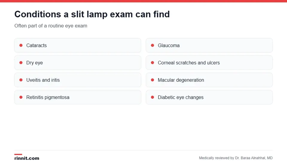
Conditions a slit lamp exam can find.
Cataracts: clouding of the lens.
Glaucoma, along with other glaucoma tests.
Dry eye and tear film problems.
Corneal abrasions, keratitis and other corneal disease.
Uveitis and iritis.
Macular degeneration and retinitis pigmentosa. Sources: [1]
You'll likely need one every few years as part of a routine eye exam, or more often if you have diabetes or an eye condition. Sources: [1]
How do I prepare?
No special preparation is needed. Because your pupils may be dilated, arrange for someone to drive you home and bring sunglasses. If you wear contact lenses, ask whether to remove them before the exam. Sources: [1]
What happens during the exam?
You sit in the exam chair and rest your chin and forehead on the supports to keep your head steady.
The specialist shines the light into and around your eyes and looks through the microscope.
They may place a yellow dye (fluorescein) in your eye with drops or a paper strip; it glows under blue light to show scratches and how quickly tears clear.
They may put in dilating drops, which take several minutes to work, then examine the back of your eye, sometimes with a handheld lens. Sources: [1]
The exam is quick and painless. The bright light may feel uncomfortable, and the drops may sting briefly. Sources: [1]
After the exam
Your eyes may be sensitive to light for a few hours while dilated; sunglasses help.
Don't drive until your pupils return to normal.
Side effects are rare and usually come from dilating drops: dizziness, dry mouth, flushed face, nausea or vomiting. Sources: [1]
Go to the emergency room if your vision suddenly worsens or you lose sight in one or both eyes after the test. Sources: [1]
Understanding your results
There's no score. Your specialist tells you what they saw and whether you need more tests or treatment, such as new glasses, eye drops, cataract surgery or referral to a retina or cornea specialist. Sources: [1, 2, 3]
Conclusion
A slit lamp exam is one of the most useful tools in eye care: a fast, painless look at nearly every part of your eye that can catch problems early. Plan for a driver if you'll be dilated, and tell your specialist about any symptoms such as pain, redness, blurred vision or flashes of light.
Frequently asked questions
What does a slit lamp exam check for?
Cataracts, glaucoma, dry eye, corneal injuries, uveitis, macular degeneration and other eye conditions.
Does a slit lamp exam hurt?
No. The light is bright, and drops may sting briefly, but it isn't painful.
How long does it take?
Only a few minutes, plus time for dilating drops to work if used.
Will my eyes be dilated?
Sometimes, to see the retina and optic nerve.
Can I drive after a slit lamp exam?
Not until your pupils return to normal if they were dilated.
What is the yellow dye for?
Fluorescein highlights scratches on the cornea and shows how well tears drain.
Who performs it?
An optometrist or ophthalmologist, usually during a routine eye exam.
How often do I need one?
Every few years, or more often with diabetes or an eye condition.
Are there side effects?
Rarely, from dilating drops: dizziness, dry mouth, flushing or nausea.
Should I remove my contacts?
Ask your specialist; lenses are often removed, especially if dye is used.
Related Reading
Cataracts: Symptoms, Causes, Diagnosis and Treatment — What You Need to Know the lens clouding a slit lamp detects.
Dry Eyes: Symptoms, Causes, Treatment & Relief — Complete Guide tear problems seen with dye on the slit lamp.
Uveitis: Symptoms, Causes, and Treatment — What Eye Inflammation Means for Your Vision eye inflammation found on a slit lamp exam.
Corneal Abrasion: Symptoms, Causes, Treatment, and Recovery corneal scratches shown with fluorescein dye.
Eye Exam: What Happens, Tests Included and How Often to Go the routine exam that includes a slit lamp exam.
References
Cleveland Clinic. Slit Lamp Exam: What It Is, Procedure & Results. Cleveland Clinic. Last updated November 1, 2022. https://my.clevelandclinic.org/health/diagnostics/24422-slit-lamp-exam
Sarayar R, Lestari YD, Setio AAA, et al. Accuracy of artificial intelligence model for infectious keratitis classification: a systematic review and meta-analysis. Front Public Health. 2023;11:1239231. PMID 38074720. https://doi.org/10.3389/fpubh.2023.1239231
Prayogo ME, Zaharo AF, Damayanti NNR, et al. Accuracy of low-cost, smartphone-based retinal photography for diabetic retinopathy screening: a systematic review. Clin Ophthalmol. 2023;17:2459-2470. PMID 37614846. https://doi.org/10.2147/OPTH.S416422
Medical disclaimer
This article is for general education only and does not replace advice from a qualified healthcare professional. Talk to your doctor about your own diagnosis and treatment, and seek emergency care right away for severe or sudden symptoms.



Comments