Corneal Topography: Mapping the Cornea and Reading the Results

Updated: 2 days ago
Written and medically reviewed by Dr. Baraa Alnahhal, MD · Last reviewed: November 2026 · How we review
Quick Answer
Corneal topography is a painless eye test that maps the shape of the cornea, the clear dome at the front of the eye, using color-coded maps. Nothing touches the eye. It helps diagnose and track astigmatism and keratoconus and is essential for planning LASIK, cataract surgery or a cornea transplant. You may need to stop wearing contacts beforehand.
What the research says
Screening matters before laser surgery: a review reported that ectasia, a bulging of the cornea after refractive surgery, occurs in about 90 per 100,000 eyes after LASIK even without known risk factors, and that undetected early keratoconus is a key risk factor that corneal mapping aims to find (Wagner FM, Sekundo W, Klin Monbl Augenheilkd, 2023) [2]
Corneal maps can be read accurately by computers: a Cochrane review of 63 studies found AI analysis of corneal topography and tomography detected manifest keratoconus with about 98.6% sensitivity and 98.3% specificity, and early (subclinical) keratoconus with 90% sensitivity, though evidence certainty was low (Vandevenne MM et al., Cochrane Database Syst Rev, 2023) [3]
Key takeaways
Maps the shape and thickness of the cornea.
Finds astigmatism and keratoconus, sometimes before symptoms.
Essential before LASIK, PRK and cataract surgery.
Quick, painless and nothing touches the eye.
You may need to stop contact lenses for weeks beforehand.
What is the cornea?
The cornea is the clear, curved front surface of your eye. It does most of the eye's focusing, so even small changes in its shape can blur vision. Corneal topography, also called computerized corneal topography, measures that shape in detail. Sources: [1]
Why is it done?

Corneal topography maps the front of the eye.
To diagnose and monitor astigmatism, keratoconus and pellucid marginal degeneration.
To check scarring from injuries or infections, and growths such as pterygium.
To plan and check results of LASIK, PRK, cataract surgery, cornea transplant and corneal cross-linking.
To fit contact lenses, especially rigid gas-permeable lenses for irregular corneas. Sources: [1]
Finding keratoconus early is especially important, because people with it have a higher risk of problems after LASIK. Sources: [1, 2]
How do I prepare?
Usually no preparation is needed. If you wear contact lenses, your eye doctor may ask you to stop for several weeks, because contacts can temporarily change the cornea's shape. Sources: [1]
What happens during the test?
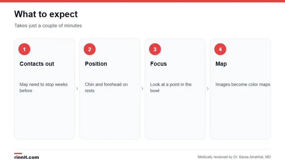
The test takes just a couple of minutes.
You sit in front of a bowl-shaped device.
You rest your chin and forehead on supports.
You look at a point inside the bowl while images are captured.
A computer turns the images into color-coded maps. Sources: [1]
The test takes just a couple of minutes and has no known risks. Sources: [1]
How to read the maps

Warm colors show steeper or thinner areas.
Map | What it shows | How to read it |
Axial (sagittal) | Curvature; the most common map | Red and orange are steeper; blue and green are flatter; normal is mostly green |
Elevation | Front and back surface height compared with a reference shape | Positive values are higher; negative values are lower |
Pachymetry | Corneal thickness | Warm colors are thinner; cool colors are thicker; keratoconus causes thinning |
Your eye doctor reviews the maps and numbers with you. Sources: [1, 3]
Conclusion
Corneal topography gives a detailed picture of your cornea's shape in minutes. It's one of the most important tests before laser vision surgery and a key tool for catching keratoconus early.
Keep learning: read about astigmatism and corneal cross-linking.
Frequently asked questions
What is corneal topography?
A test that maps the shape of the cornea.
Does it hurt?
No. Nothing touches your eye.
How long does it take?
Just a couple of minutes.
Why stop contact lenses first?
Contacts can temporarily change the cornea's shape.
What can it detect?
Astigmatism, keratoconus, scarring and growths.
Why is it done before LASIK?
To find corneal problems that raise the risk of complications.
What do red areas mean?
Steeper areas on the curvature map, or thinner areas on the thickness map.
What does a normal map look like?
Mostly green on the curvature map.
Is it the same as keratometry?
No. Topography maps the whole surface; keratometry measures a few points.
Are there risks?
No known risks.
Related Reading
Corneal Pachymetry: Measuring Corneal Thickness for Glaucoma and LASIK explains the test that measures corneal thickness.
Understanding the Cornea: The Eye’s Protective Shield the part of the eye this test maps.
Astigmatism: Symptoms, Causes & Treatment Options — Complete Guide a common finding on corneal maps.
Corneal Cross-Linking (CXL): Purpose, Procedure, Risks and Recovery a treatment for keratoconus.
Photorefractive Keratectomy (PRK) Eye Surgery laser surgery planned with topography.
Eye Exam: What Happens, Tests Included and How Often to Go where eye tests begin.
Keratometry: Measuring Corneal Curve for Astigmatism and Cataract Surgery explains the test that measures the curve of the cornea.
References
Cleveland Clinic. Corneal Topography: What To Expect & How To Interpret Results. Cleveland Clinic. Last updated March 13, 2023. https://my.clevelandclinic.org/health/diagnostics/24819-corneal-topography
Wagner FM, Sekundo W. Iatrogenic keratectasia after refractive surgery: causes, prophylaxis, therapy. Klin Monbl Augenheilkd. 2023;240(6):783-794. PMID 37348513. https://doi.org/10.1055/a-2073-8478
Vandevenne MM, Favuzza E, Veta M, et al. Artificial intelligence for detecting keratoconus. Cochrane Database Syst Rev. 2023;11(11):CD014911. PMID 37965960. https://doi.org/10.1002/14651858.CD014911.pub2
Medical disclaimer
This article is for general education only and does not replace advice from a qualified healthcare professional. Talk to your doctor about your own diagnosis and treatment, and seek emergency care right away for severe or sudden symptoms.



Comments