Carotid Ultrasound: Why It's Done, What to Expect and What Results Mean

Updated: 2 days ago
Written and medically reviewed by Dr. Baraa Alnahhal, MD · Last reviewed: November 2026 · How we review
Quick Answer: What is a carotid ultrasound?
A carotid ultrasound (carotid duplex) is a painless test that uses sound waves to make pictures of the two carotid arteries in the neck, which carry blood to the brain. It looks for plaque, blood clots and narrowing that can raise the risk of stroke. The test takes 30 to 45 minutes, needs no preparation and uses no radiation. [1]
What the research says
Duplex ultrasound is accurate at telling whether significant narrowing is present. A Cochrane review of 22 studies (4,957 carotid arteries) found duplex ultrasound had a summary sensitivity of 97% and specificity of 70% for 50% to 99% narrowing, and 85% sensitivity for 70% to 99% narrowing. The authors advise caution when using it as the only test before surgery. (Cassola et al., Cochrane Database of Systematic Reviews, 2022) [2]
Speed cutoffs work, but results vary by lab. A meta-analysis found that a peak blood speed of 200 cm/s or more detected narrowing of 70% or more with 90% sensitivity and 94% specificity. Accuracy varied widely between laboratories, which matters for treatment decisions. (Jahromi et al., Journal of Vascular Surgery, 2005) [3]
Key takeaways
A carotid ultrasound checks the arteries in your neck for narrowing or blockage.
It is painless, uses sound waves instead of radiation and has no known risks.
You don't need to prepare; just wear a shirt that leaves your neck open.
Results show how blocked an artery is, as a percentage.
Treatment ranges from lifestyle changes and medicine to a procedure, depending on how severe the blockage is.
What is a carotid ultrasound?
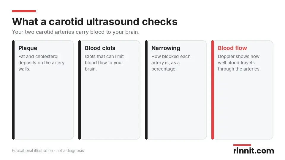
A carotid ultrasound, also called a carotid duplex, uses sound waves to show what your carotid arteries look like on the inside. You have one carotid artery on each side of your neck, and together they carry blood to your brain. [1]
Using a Doppler technique, your provider can also see how well blood travels through these arteries. A sonographer (ultrasound technologist) or a radiologist performs the test. [1]
Why would you need a carotid ultrasound?
Your provider may order this test to look for blood clots or plaque (fat and cholesterol deposits) on the walls of your carotid arteries. Plaque can limit, and eventually block, blood flow to your brain, face and neck. A blocked carotid artery can cause a stroke. Call 911 right away if you have stroke signs such as face drooping, arm or leg weakness, or trouble speaking. [1]
You may need a carotid ultrasound if you: [1]
Had surgery on a narrowed artery or need a follow-up on a carotid stent
Are being checked regularly because an artery was narrow before
Have a bruit, an unusual whooshing sound your provider hears with a stethoscope
Have high blood pressure, high cholesterol or diabetes
Had a stroke or transient ischemic attack (TIA)
Are about to have coronary artery bypass surgery
Have stroke or heart disease in your family
Have a hematoma (a pool of blood)
Doppler ultrasound helps your provider look at blood flow, narrowing or blockage, and malformations present since birth or tumors. [1]
How does a carotid ultrasound work?
The test doesn't use radiation. A handheld device called a transducer sends sound waves into your neck. The waves bounce off your arteries and return to the transducer, and a computer turns them into images on a monitor. The sonographer can save still images or record video. [1]
How do you prepare?
You don't need to do anything to prepare. Wear a comfortable shirt that doesn't cover your neck and isn't tight, or you may need to change into a hospital gown. Skip necklaces or earrings that might get in the way. [1]
What happens during the test?
The test is done at your provider's office or a healthcare facility and takes about 30 to 45 minutes. [1]
You lie on your back.
Your provider puts clear gel on both sides of your neck. It may feel warm and helps sound waves travel.
They press the transducer, which is smaller than a store price scanner, against your skin.
They move it over your neck until they have all the images they need.
You stay still so the images are clear.
You won't hear the sound waves unless Doppler is used. Then you may hear swishing sounds that change pitch. [1]

After the test
You'll get a towel to wipe off the gel. Then you can get dressed and go home, and return to your normal activities. [1]
Are there risks?
This test has no risks. You may feel pressure, but not pain, when the transducer presses on your neck. [1]
What do carotid ultrasound results mean?
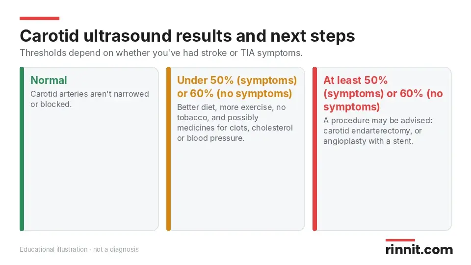
Your result tells whether your arteries are blocked and by how much, given as a percentage. A normal result means your carotid arteries aren't narrowed or blocked. An abnormal result may mean you have atherosclerosis, a blood clot or another problem that narrows the artery and raises your stroke risk. [1]
Result | Treatment your provider may suggest |
Normal | No narrowing or blockage |
Plaque under 50% (with stroke or TIA symptoms) or under 60% (without symptoms) | Better diet, more exercise, stopping tobacco, and possibly medicines |
Plaque of at least 50% (with symptoms) or at least 60% (without symptoms) | A procedure such as carotid endarterectomy, or angioplasty with a stent |
Sources: [1]
Medicines your provider may prescribe
Thrombolytics to dissolve blood clots
Aspirin or clopidogrel to prevent blood clots
Statins to lower cholesterol
Antihypertensives to lower blood pressure
Sources: [1]
Procedures for severe blockage
A carotid endarterectomy removes the plaque, and the ultrasound helps plan it by showing where the blockage is. Angioplasty pushes plaque against the artery walls to make more room for blood, and a stent can hold the artery open afterward. [1]
How accurate is a carotid ultrasound?
It is generally accurate, but sometimes it looks like there is a blockage when there isn't. Sonographer experience varies, and ultrasound picks up severe problems better than mild ones. Bone can also block the view of part of the artery. Your provider may order more imaging, such as CT angiography, magnetic resonance angiography or cerebral angiography. [1]
What raises your stroke risk?
Other risk factors can affect which treatment your provider recommends. You may be at higher risk of stroke if you have: [1]
High blood pressure or high cholesterol
Diabetes
Peripheral artery disease (PAD)
Atrial fibrillation
Sickle cell disease
Obesity or heart disease
Using tobacco, eating a high-fat, high-salt diet and being inactive also raise stroke risk. [1]
When will you get your results?
Sometimes a radiologist talks with you right away. Other times, the results go to your provider, who shares them in a couple of days. If you haven't heard back after a few days, contact your provider. [1]
Questions to ask your provider
How much narrowing do I have in each carotid artery?
Do I need more imaging to confirm the result?
Which treatment is right for my level of blockage?
What lifestyle changes will lower my stroke risk most?
How often should I repeat this test?
Conclusion
A carotid ultrasound is a quick, painless way to find narrowing or blockage in the arteries that supply your brain. Its results, given as a percentage of blockage, guide whether you need lifestyle changes, medicines or a procedure.
Even if you need treatment, healthier habits like not smoking, staying active and eating well can lower your stroke risk further.
Keep learning: Read our guides to Doppler ultrasound, ultrasound, the angiogram and the lipid panel. For more on this test, see MedlinePlus: Carotid duplex.
FAQ
What does a carotid ultrasound check for?
It checks your neck arteries for plaque, blood clots, narrowing and how well blood flows to your brain. [1]
Is a carotid ultrasound painful?
No. You may feel pressure from the transducer, but it doesn't hurt. [1]
How long does a carotid ultrasound take?
It usually takes about 30 to 45 minutes. [1]
Do I need to prepare for a carotid ultrasound?
No. Just wear a comfortable shirt that leaves your neck open and skip necklaces or earrings. [1]
Does a carotid ultrasound use radiation?
No. It uses sound waves, not radiation. [1]
Are there risks?
This test has no risks. [1]
What is a normal carotid ultrasound result?
A normal result means your carotid arteries aren't narrowed or blocked. [1]
What happens if my artery is blocked?
Mild blockage is treated with lifestyle changes and possibly medicines; severe blockage may need endarterectomy or angioplasty with a stent. [1]
How accurate is a carotid ultrasound?
It is generally accurate, but it may suggest a blockage that isn't there, so more imaging is sometimes needed. [1]
When will I get my results?
Sometimes right away from a radiologist, or within a couple of days from your provider. [1]
Related Reading
Carotid Artery Stenting: Procedure, Recovery and Results explains carotid artery stenting.
Carotid Endarterectomy: Surgery, Risks and Recovery explains carotid artery surgery to prevent stroke.
Carotid Intima-Media Thickness (CIMT) Test: Checking Artery Walls explains the ultrasound that measures carotid artery wall thickness.
Carotid Angiography: Imaging Neck Arteries for Stroke Risk explains the X-ray dye test that shows narrowing in the neck arteries.
Learn more in our guide to Carotid Artery Disease: The Silent Stroke Risk in Your Neck, Explained.
Learn more in our guide to Stroke: Symptoms, Causes, Diagnosis, and Treatment — A Complete Guide.
Learn more in our guide to Transient Ischemic Attack (TIA): The Mini-Stroke That Is Actually a Warning.
Learn more in our guide to Arteriosclerosis & Atherosclerosis: Symptoms, Causes, Diagnosis & Treatment — Complete Guide.
Learn more in our guide to Understanding Hyperlipidemia: Managing High Cholesterol for Heart Health.
Learn more in our guide to Carotid Artery: Anatomy, Function, and Stroke Prevention.
Vascular Ultrasound: How It Checks Blood Flow, What It Finds and What to Expect explains the ultrasound that checks blood flow in arteries and veins.
References
Cleveland Clinic. Carotid Ultrasound: Purpose, Procedure and Results. Cleveland Clinic. Last updated April 29, 2022. https://my.clevelandclinic.org/health/diagnostics/22916-carotid-ultrasound
Cassola N, Baptista-Silva JC, Nakano LC, et al. Duplex ultrasound for diagnosing symptomatic carotid stenosis in the extracranial segments. Cochrane Database Syst Rev. 2022;7(7):CD013172. PMID 35815652. https://doi.org/10.1002/14651858.CD013172.pub2
Jahromi AS, Cinà CS, Liu Y, et al. Sensitivity and specificity of color duplex ultrasound measurement in the estimation of internal carotid artery stenosis: a systematic review and meta-analysis. J Vasc Surg. 2005;41(6):962-972. PMID 15944595. https://doi.org/10.1016/j.jvs.2005.02.044
Medical disclaimer
This article is for general education only and does not replace advice from a qualified healthcare professional. Talk to your doctor about your own diagnosis and treatment, and seek emergency care right away for severe or sudden symptoms.



Comments