Carotid Angiography: Imaging Neck Arteries for Stroke Risk

Updated: 3 days ago
Written and medically reviewed by Dr. Baraa Alnahhal, MD · Last reviewed: November 2026 · How we review
Quick Answer
Carotid angiography is an imaging test that uses X-rays and contrast dye to show the carotid arteries, the main blood vessels in the neck that carry blood to the brain. It reveals narrowing or blockages. It's often done after a TIA or stroke, or when a carotid ultrasound is unclear. The test takes 60 to 90 minutes.
What the research says
Finding severe narrowing changes outcomes: in 659 people with recent symptoms and 70% to 99% carotid narrowing on angiography, the 2-year risk of stroke was 26% with medicine alone vs. 9% after carotid endarterectomy (NASCET Collaborators, New England Journal of Medicine, 1991) [2]
Serious complications are rare: in 19,826 diagnostic angiograms of the brain and neck arteries, neurological complications occurred in 2.63%, strokes with permanent disability in 0.14% and a bruise-like collection of blood at the access site (hematoma) was the most common problem (Kaufmann TJ et al., Radiology, 2007) [3]
Key takeaways
Carotid angiography shows narrowing and blockages in the neck arteries.
A catheter and contrast dye are used with X-rays.
It takes 60 to 90 minutes with sedation and local numbing.
You rest for several hours afterward and drink fluids to flush the dye.
Results guide treatment to lower stroke risk.
What is carotid angiography?
The carotid arteries can narrow when plaque builds up inside them, which raises the risk of stroke. An angiogram gives detailed pictures of the inside of these arteries. An interventional radiologist performs the catheter-based test. Less invasive versions use a CT angiogram or an MRA. Sources: [1]
Types of carotid angiography

Your provider chooses the test that fits your situation.
Type | How it works | Notes |
Catheter angiography | Catheter delivers dye directly; X-ray pictures | Most detailed; invasive |
CT angiography | IV dye with a CT scan | Noninvasive; uses radiation |
MR angiography | MRI, often with IV contrast | Noninvasive; no radiation |
Sources: [1]
Why it's done
To check for suspected carotid artery disease.
After a TIA or stroke.
When a carotid ultrasound result is unclear.
To plan treatment, such as endarterectomy or stenting. Sources: [1]
How to prepare
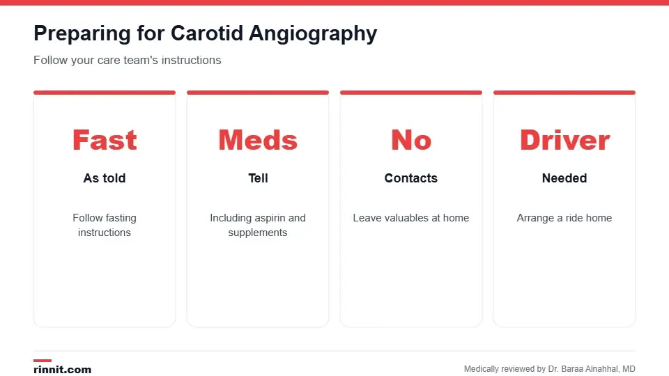
Your care team will give you exact instructions.
Follow your fasting instructions.
Tell your provider about all medicines, including aspirin and blood thinners, and herbal supplements, and follow their instructions.
Leave valuables at home and don't wear contact lenses.
Arrange for someone to drive you home. Sources: [1]
What happens during the test

The test takes 60 to 90 minutes.
An IV is placed and you receive a sedative to relax; EKG electrodes are attached.
The skin at the insertion site, usually the groin or sometimes the arm, is numbed.
A catheter is guided through the blood vessels to the carotid arteries.
Contrast dye is injected while X-ray pictures are taken.
The catheter is removed and a bandage is applied. Sources: [1]
You may feel brief warmth when the dye is injected. Tell the team right away about chest pain, itching, nausea, throat tightness or vision changes. Sources: [1]
Recovery and risks
You rest for several hours while your vital signs and the insertion site are checked. Drink plenty of fluids to flush out the dye. Possible risks include bleeding or bruising at the insertion site, allergic reaction to the dye and, rarely, stroke or other neurological problems. Call your provider for signs of an allergic reaction or if you feel unwell. Sources: [1, 3]
Results
A radiologist reviews the images and sends a report to your provider. Significant narrowing can mean a higher stroke risk. Depending on the results, treatment may include carotid endarterectomy, carotid stenting or medicines to lower clot risk. Sources: [1, 2]
Conclusion
Carotid angiography gives a clear, detailed view of the arteries that feed your brain. It helps your care team judge your stroke risk and choose the right treatment, especially after a TIA or stroke.
Keep learning: read about carotid artery disease and the carotid ultrasound.
Frequently asked questions
What is carotid angiography?
An X-ray test with dye that shows the arteries in your neck.
Why is it done?
To find narrowing or blockages that raise stroke risk.
How long does it take?
About 60 to 90 minutes.
Will I be awake?
Usually yes, with a sedative and local numbing.
Does it hurt?
It's usually painless, though the dye may feel warm.
What are the risks?
Bruising, dye allergy and, rarely, stroke.
How long is recovery?
You rest for several hours before going home.
Are there noninvasive options?
Yes. CT angiography and MR angiography.
What happens if a blockage is found?
Treatment may include endarterectomy, stenting or medicine.
Can I drive home?
No. Arrange for someone to drive you.
Related Reading
Carotid Intima-Media Thickness (CIMT) Test: Checking Artery Walls explains the ultrasound that measures carotid artery wall thickness.
Carotid Ultrasound: Why It's Done, What to Expect and What Results Mean the first test usually used to check the carotid arteries.
Carotid Artery Disease: The Silent Stroke Risk in Your Neck, Explained the condition carotid angiography evaluates.
Cerebral Angiogram: What It Shows, How to Prepare, What to Expect, Risks and Recovery an angiogram of the brain's blood vessels.
Endarterectomy: Procedure, Types, Risks and Recovery Explained surgery to remove carotid plaque.
Transient Ischemic Attack (TIA): The Mini-Stroke That Is Actually a Warning a warning sign that may lead to this test.
References
Cleveland Clinic. Carotid Angiography. Cleveland Clinic. Last updated May 2, 2022. https://my.clevelandclinic.org/health/diagnostics/16842-carotid-angiography
North American Symptomatic Carotid Endarterectomy Trial Collaborators, Barnett HJM, Taylor DW, et al. Beneficial effect of carotid endarterectomy in symptomatic patients with high-grade carotid stenosis. New England Journal of Medicine. 1991;325(7):445-453. PMID 1852179. https://doi.org/10.1056/NEJM199108153250701
Kaufmann TJ, Huston J, Mandrekar JN, et al. Complications of diagnostic cerebral angiography: evaluation of 19,826 consecutive patients. Radiology. 2007;243(3):812-819. PMID 17517935. https://doi.org/10.1148/radiol.2433060536
Medical disclaimer
This article is for general education only and does not replace advice from a qualified healthcare professional. Talk to your doctor about your own diagnosis and treatment, and seek emergency care right away for severe or sudden symptoms.



Comments