Cerebral Angiogram: What It Shows, How to Prepare, What to Expect, Risks and Recovery

Updated: 2 days ago
Written and medically reviewed by Dr. Baraa Alnahhal, MD · Last reviewed: November 2026 · How we review
Quick Answer: What is a cerebral angiogram?
A cerebral angiogram is a minimally invasive test that shows problems with the blood vessels in and around the brain. A specialist passes a catheter through an artery, injects contrast dye and takes X-rays. It can find brain aneurysms, arteriovenous malformations and narrowed or blocked arteries. Serious problems are rare; stroke happens in fewer than 1% of cases.
What the research says
Serious complications of diagnostic cerebral angiography are uncommon. In 19,826 consecutive patients, neurologic complications occurred in 2.63% of exams, strokes with permanent disability in 0.14% and deaths in 0.06%, while a bruise or blood collection at the access site was the most common problem at 4.2%. (Kaufmann et al., Radiology, 2007) [2]
Noninvasive MR angiography finds most aneurysms, but it is not as definitive. A meta-analysis including 960 patients found MR angiography detected intracranial aneurysms with a pooled sensitivity of 95% and specificity of 89%, with missed or false findings mainly near the skull base and middle cerebral artery. (Sailer et al., Stroke, 2014) [3]
Key takeaways
A cerebral angiogram uses a catheter, contrast dye and X-rays to show the brain's blood vessels [1].
It gives much more detailed pictures than CT or MRI and is also called digital subtraction angiography (DSA) [1].
It helps diagnose brain aneurysms, arteriovenous malformations, vasculitis, artery tears and clot-related stroke [1].
You may need to fast for four to eight hours if you are sedated, and someone must drive you home [1].
The test usually takes one to three hours, followed by a few hours of observation [1].
Rest for eight to 12 hours afterward and seek help right away for weakness, numbness, slurred speech or vision changes [1].
What is a cerebral angiogram?
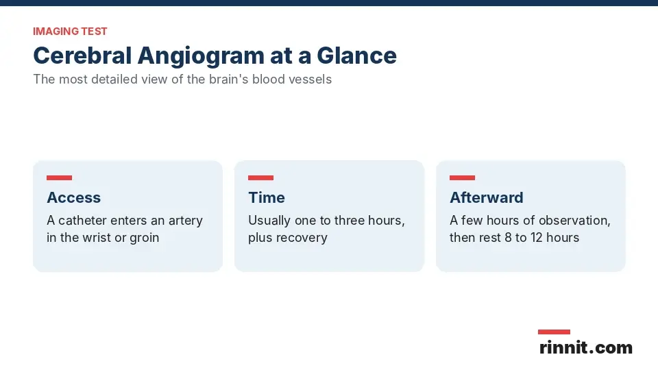
A cerebral angiogram is a diagnostic procedure that shows problems with the blood vessels in your brain [1]. A specially trained provider inserts a catheter, a thin plastic tube, into an artery in your wrist or groin and injects contrast dye through it [1]. X-rays of the blood vessels are taken while you lie on a table [1].
The test gives much more detailed images than a CT scan or MRI [1]. It is also called digital subtraction angiography of the brain, or DSA [1].
Why you might need a cerebral angiogram
Your provider may order a cerebral angiogram to diagnose or confirm a problem with the blood vessels in your brain, such as [1]:
It can also be used to check the arteries of the head and neck before treatment or surgery, to see how blood vessels feed a brain tumor and to look more closely at something unusual found on a CT or MRI [1].
How to prepare for a cerebral angiogram
Your provider will check that the test is safe for you with a physical exam, blood tests and a review of your medical history [1]. Tell them about [1]:
If you will be sedated, you may need to stop eating and drinking for four to eight hours beforehand [1]. Anyone who has sedation or general anesthesia needs someone to drive them home [1]. Follow your provider's instructions about medicines and food [1].
What happens during a cerebral angiogram
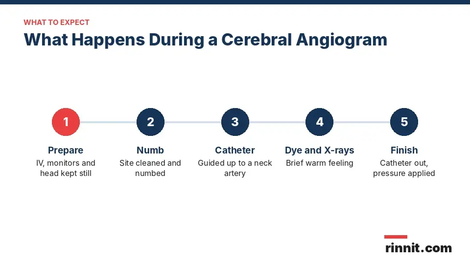
The main steps are [1]:
You change into a gown and an IV line is placed in your hand or arm for sedation if needed. Children and teens may need general anesthesia [1].
Monitors track your heart rate and blood pressure while you lie on the exam table, and your head is kept still with a strap, tape or foam holder [1].
The catheter site, usually the groin, leg or arm, is cleaned and numbed with local anesthetic, which may briefly burn or sting [1].
A very small cut is made, and the catheter enters an artery through a tiny needle hole under imaging guidance. You may feel some pressure [1].
The catheter is guided up to an artery in the neck, which you do not feel [1].
Contrast dye is injected, causing a brief warm feeling, and several sets of X-rays are taken. You need to stay very still and may be asked to hold your breath [1].
The catheter is removed and pressure is applied to prevent bleeding under the skin. A closure device may be used, and a bandage covers the site [1].
How long does it take?
The procedure usually takes one to three hours, plus extra time for preparation and recovery [1].
Is a cerebral angiogram surgery?
No. It is a minimally invasive diagnostic procedure. Although it uses a small cut to place the catheter, it does not change any body tissue [1].
Recovery after a cerebral angiogram
You stay in a recovery room for a few hours of observation and then go home [1]. You can eat normally right away, but you should rest for eight to 12 hours before going back to other activities [1]. If you are breastfeeding, you can usually keep breastfeeding after the contrast injection; if you prefer, you can pause and pump for 12 to 24 hours [1].
Risks of a cerebral angiogram
As with any catheter procedure, risks include damage to a blood vessel with possible internal bleeding, bruising or bleeding at the puncture site and infection [1]. Other risks include [1]:
Risk | What to know |
Allergic reaction to contrast | Rare; the radiologist can treat it [1] |
Kidney damage from contrast | Mainly in people with diabetes or kidney disease; the kidneys usually recover within five to seven days [1] |
Blood clot at the catheter tip | Rare; may need surgery to reopen the vessel [1] |
Stroke | Can happen if the catheter loosens plaque; occurs in fewer than 1% of cases [1] |
Sources: [1]
Cerebral angiogram results
A radiologist writes a report and sends it to the provider who ordered the test, who will discuss the results with you [1]. You may need follow-up tests [1].
What the images show | What it may mean |
Contrast leaking out of a vessel | Bleeding [1] |
Narrowed or blocked arteries | Plaque, artery spasm (vasospasm), inherited disorders or a clot causing a stroke [1] |
Blood vessels out of their usual place | A brain tumor, bleeding inside the skull, an aneurysm or an arteriovenous malformation [1] |
Sources: [1]
When to get help right away
Call your provider or seek emergency care right away if you have any of these after the test [1]:

Conclusion
A cerebral angiogram gives the most detailed view of the blood vessels in and around the brain, which helps diagnose aneurysms, AVMs, narrowed arteries and other vessel problems. It is a minimally invasive test with a short stay for observation afterward.
Serious complications are rare. Rest for the rest of the day, watch the catheter site and get help right away for any signs of stroke.
Keep learning: Read our guide to the CT angiogram (CTA), our overview of the angiogram or our guide to the carotid ultrasound. For more background, see the RadiologyInfo guide to cerebral angiography.
FAQ
What is a cerebral angiogram?
It is a test that uses a catheter, contrast dye and X-rays to show the blood vessels in and around the brain.
What can a cerebral angiogram detect?
It can find brain aneurysms, AVMs, dural fistulas, vasculitis, artery tears, narrowed arteries and clots causing stroke.
Is a cerebral angiogram better than a CT or MRI?
It gives much more detailed pictures of the brain's blood vessels than CT or MRI.
Where is the catheter inserted?
Usually through an artery in the wrist or groin, then guided up to an artery in the neck.
Do I need to fast before a cerebral angiogram?
If you are sedated, you may need to fast for four to eight hours. Follow your provider's instructions.
Does a cerebral angiogram hurt?
The numbing medicine may sting briefly, you may feel pressure and the dye causes a short warm feeling.
How long does a cerebral angiogram take?
Usually one to three hours, plus time to prepare and recover.
What are the risks of a cerebral angiogram?
Bruising, bleeding, infection, dye allergy, kidney strain, clots and, in fewer than 1% of cases, stroke.
When can I go back to normal activities?
You can eat right away but should rest for eight to 12 hours before other activities.
When should I get help after a cerebral angiogram?
Right away for weakness, numbness, slurred speech, vision changes, chest pain, trouble breathing or signs of infection at the site.
Related Reading
Carotid Angiography: Imaging Neck Arteries for Stroke Risk explains the X-ray dye test that shows narrowing in the neck arteries.
Brain Aneurysm: Symptoms, Causes, and Treatment explains brain aneurysms, one of the main problems a cerebral angiogram is used to confirm.
Brain AVM (Arteriovenous Malformation): Symptoms, Causes, and Treatment covers arteriovenous malformations, tangled vessels this test can map in detail.
Dural Arteriovenous Fistulas (DAVF): What They Are, Symptoms, and Treatment Options explains abnormal artery-to-vein connections that a cerebral angiogram can diagnose.
Vasculitis: Symptoms, Causes, Treatment & Complete Guide covers inflamed blood vessels, which can show up as narrowing on the images.
What Is Moyamoya Disease? A Plain-Language Guide to This Rare Brain Blood Vessel Condition explains a rare narrowing of brain arteries that angiography helps confirm.
Subarachnoid Hemorrhage (SAH): Symptoms, Causes, and Emergency Care covers bleeding around the brain, often investigated with a cerebral angiogram.
Wada Test: Finding Language and Memory Before Brain Surgery explains the test that shows which side of the brain controls language and memory.
References
Cleveland Clinic. Cerebral Angiogram: What It Is, Purpose, Procedure & Risks. Cleveland Clinic. Last updated August 8, 2023. https://my.clevelandclinic.org/health/diagnostics/13476-cerebral-angiogram
Kaufmann TJ, Huston J, Mandrekar JN, et al. Complications of diagnostic cerebral angiography: evaluation of 19,826 consecutive patients. Radiology. 2007;243(3):812-819. PMID 17517935. https://doi.org/10.1148/radiol.2433060536
Sailer AM, Wagemans BA, Nelemans PJ, et al. Diagnosing intracranial aneurysms with MR angiography: systematic review and meta-analysis. Stroke. 2014;45(1):119-126. PMID 24326447. https://doi.org/10.1161/STROKEAHA.113.003133
Medical disclaimer
This article is for general education only and does not replace advice from a qualified healthcare professional. Talk to your doctor about your own diagnosis and treatment, and seek emergency care right away for severe or sudden symptoms.



Comments