Dural Arteriovenous Fistulas (DAVF): What They Are, Symptoms, and Treatment Options

Updated: 3 days ago
Medically reviewed by Dr. Baraa Alnahhal, MD · Last reviewed: October 2026
A dural arteriovenous fistula (dAVF) is a rare, abnormal connection between blood vessels in the dura mater, the brain's outer covering [1]. It can lead to bleeding in the brain or other serious symptoms [1] and tends to occur between ages 50 and 60 [1]. Symptoms range from none at all to aggressive problems such as sudden headache, seizures and pulsatile tinnitus, which means hearing a rhythmic sound in the ear that matches your heartbeat [1]. Treatment involves blocking or disconnecting the fistula with an endovascular procedure, focused radiation or surgery [2].
Quick Answer: What is a dural arteriovenous fistula (DAVF), and how is it treated?
A dural arteriovenous fistula (DAVF) is a rare, abnormal connection between blood vessels in the dura mater, the brain's outer covering, that can lead to brain bleeding. It most often affects people between ages 50 and 60. Symptoms vary from none to sudden headache, seizures or pulsatile tinnitus. Diagnosis uses angiography, and treatment blocks or disconnects the fistula. [1] [2]
What the research says
Endovascular embolization closes most fistulas it targets. A meta-analysis of 10 studies (214 patients, 113 of them with dAVFs) found complete occlusion in 91% of dAVFs treated with the newer liquid embolic agents Squid or PHIL, with a permanent procedure-related morbidity rate of 3% (for example, Günkan et al., Interventional Neuroradiology, 2024 [3]).
Fistulas near the eye socket can be treated with low risk. Across 22 studies and 1,043 patients with cavernous sinus dAVFs, 85% had a complete resolution of symptoms after endovascular treatment, complications occurred in 7.75%, and there was one death (Alexandre et al., Clinical Neuroradiology, 2021 [4]).
Fistulas at the front of the skull base carry a high bleeding risk but respond well to treatment. In 18 studies of 231 anterior cranial fossa dAVFs, endovascular treatment achieved complete occlusion in 85%, symptoms resolved or improved in 94% of successfully embolized patients, and morbidity and mortality were 1% and 0% (Ferreira et al., Neurosurgical Review, 2024 [5]).
What Is a Dural Arteriovenous Fistula?
A dural arteriovenous fistula (dAVF) is an unusual connection between blood vessels in the brain's outer covering, which is called the dura mater [1]. These abnormal connections can lead to bleeding in the brain or other serious symptoms [1].
Dural AVFs are rare and tend to occur between ages 50 and 60 [1]. Two facts tend to reassure people with a family history. First, dural AVFs are not typically genetic, so children are not more likely to develop a dAVF if their parent has one [1]. Second, some people with dAVFs have no symptoms at all [1].

Symptoms: From Silent to Serious
Symptoms of dural arteriovenous fistulas can be different for each person, depending on where the dAVF is located. Some people with dAVFs may not have symptoms. When symptoms occur, they may be characterized as benign or aggressive, and an aggressive dAVF has more serious symptoms [1].
Aggressive Symptoms
Aggressive dAVF symptoms can result from bleeding in the brain, known as intracerebral hemorrhage. Bleeding in the brain often causes a sudden headache, along with other symptoms based on the location and size of the hemorrhage [1].
Aggressive symptoms can also result from nonhemorrhagic neurological deficits (NHNDs), which are problems that happen without bleeding. These can include seizures or changes in thinking and memory, and they usually develop more gradually, over days to weeks. Symptoms are typically related to the area of the brain affected [1].
Aggressive symptoms can include [1]:
Sudden headache
Trouble walking and falls
Seizures
Speech or language issues
Facial pain
Dementia, which rarely may occur due to increased pressure in the blood vessels in the brain
Parkinsonism: slowed movement, stiffness and tremor
Trouble with coordination
Burning or prickling sensations
Weakness
Apathy: lack of interest
Failure to thrive
Pressure-related symptoms: headaches, nausea and vomiting
Hearing and Vision Symptoms

Other dAVF symptoms can involve hearing and vision. People with hearing symptoms may experience pulsatile tinnitus: hearing a rhythmic sound in the ear that occurs with the heartbeat [1].
Vision-related symptoms may include [1]:
Vision changes
An eye bulge
Swelling in the eye lining
Paralysis of a muscle in or around the eye
When to See a Doctor
Make an appointment with your healthcare professional if you have any symptoms that are not usual or that worry you [1].
Seek medical help immediately if you experience a seizure or symptoms that suggest brain hemorrhage, such as [1]:
Sudden, severe headache
Nausea and vomiting
Weakness or numbness on one side of the body
Trouble speaking or understanding speech
Loss of vision or double vision
Trouble with balance
A sudden severe headache combined with any of these signs is a medical emergency. Don't wait it out.
What Causes a DAVF?
It's not clear why dural arteriovenous fistulas happen. But some things that might cause them include [1]:
Traumatic head injury
Infection
Previous brain surgery
Blood clots in deep veins
Tumors
Most experts think that dAVFs involving larger brain veins form when one of the brain's venous sinuses narrows or becomes blocked. The venous sinuses are channels in the brain that route circulated blood from the brain back to the heart [1].
Who Is Most at Risk?
A key risk factor is being inclined to blood clots in veins, known as vein thrombosis. Changes in the way blood clots may increase the risk of a blockage or narrowing of the venous sinuses [1].
In terms of age, dAVFs most often affect people between 50 and 60, but they can occur at younger ages, including in children. Research has also found that noncancerous tumors in the membranes that surround the brain and spinal cord may be associated with dAVFs [1].
Can you prevent a DAVF? Because the exact cause isn't known, there's no one way to prevent them from happening [1].
How a DAVF Is Diagnosed
If you have symptoms of a dural arteriovenous fistula, you may need imaging tests [2]:
Digital subtraction angiography (DSA). The most reliable tool to diagnose dAVF. It takes very clear pictures of blood flow and is essential for defining how many fistulae exist and where, the anatomy of the external carotid arteries and any branches between them and the dura, the structure of the fistula's blood vessels, whether cardiovascular disease is also present, how much narrowing or blockage has occurred in the dural sinus, and whether any affected veins are dilated and to what extent.
Time-resolved CT angiography (CTA). Can help your healthcare professional see if the arteries and veins are connected incorrectly or if there's any unusual blood flow.
Magnetic resonance angiography (MRA). A special type of MRI used to watch how blood flows through your brain. A dye may be used to highlight blood vessels and create more-detailed images.
Magnetic resonance imaging (MRI). Can reveal the shape of a dAVF and detect very small bleeds, and may determine the impact of any irregular blood vessel structures.
CT scans. Can show fluid buildup caused by increased blood pressure in the brain, and bleeding that may be caused by a dAVF.
Treatment: How a DAVF Is Fixed
Treatment for a dural arteriovenous fistula involves a procedure to block or disconnect the fistula [2]. Options include:
Endovascular procedures. A long, thin tube called a catheter is inserted into a blood vessel in your leg or groin and threaded through blood vessels to the fistula using X-ray imaging. Coils or a glue-like substance is released to block the connection in the blood vessels.
Stereotactic radiosurgery. Precisely focused radiation blocks the irregular connection in the blood vessels, which causes the blood vessels in the fistula to close off and destroys the dAVF. Technologies used include the linear accelerator, Gamma Knife, CyberKnife and proton beam therapy.
dAVF surgery. If an endovascular procedure or stereotactic radiosurgery aren't options, surgery may be performed to disconnect the dAVF or cut off the blood supply and remove the fistula.
Sometimes a combination of approaches is recommended. For example, endovascular embolization may be performed shortly after radiosurgery, which can help speed up symptom relief and provide long-term results [2].
Preparing for Your Appointment
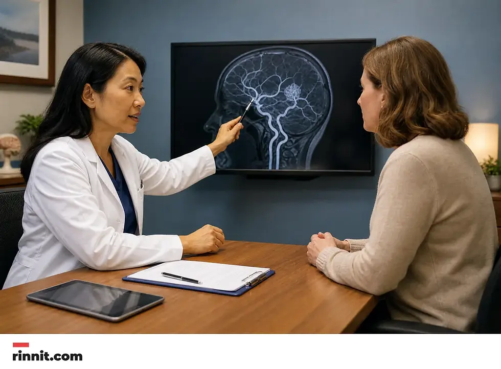
You'll likely start with a specialist consultation, often with a neurologist. Coming prepared makes the visit far more productive [2].
What You Can Do
Keep a detailed symptoms calendar. Each time a symptom occurs, write down the date and time, what you experienced and how long it lasted.
Write down key personal information, including any major stresses or recent life changes.
Make a list of all medicines, vitamins and supplements you're taking, with doses.
Take a family member or friend along. Someone with you may remember something that you missed or forgot.
Bring any recent brain scans on a CD. If you've had seizures, your healthcare professional may want to ask questions of someone who has witnessed them, because it's common not to be aware of everything that happens during a seizure.
Questions Worth Asking
Write your questions down and rank them from most to least important, in case time runs out. Useful ones include [2]:
Where is the fistula located?
Will I need follow-up tests, and how often?
How long would surgery take, and how long is recovery?
How long should I plan to be in the hospital?
How many dAVFs have you seen and how many have you treated?
Does your institution have a cerebrovascular specialty practice?
In the Meantime
Certain conditions and activities can trigger seizures, so it may help to avoid drinking excessive amounts of alcohol, avoid nicotine, get enough sleep and reduce stress [2].
Bottom Line
A dural arteriovenous fistula is a rare condition that ranges from silent to serious depending on its location and behavior [1]. Treatment is available: the abnormal connection can be blocked or removed through endovascular procedures, focused radiation or surgery, sometimes in combination [2].
The watchwords are awareness and speed. Unusual symptoms, especially a sudden severe headache, pulsatile tinnitus, seizures or new weakness, deserve prompt medical attention. A clear diagnosis and a specialist team are the fastest path to effective treatment.
Your next step: if a symptom described here matches something you are experiencing, make an appointment with your healthcare professional. If it is a seizure or a sudden severe headache with other warning signs, seek emergency care immediately.
Frequently Asked Questions
What is a dural arteriovenous fistula?
A dural arteriovenous fistula (DAVF) is a rare, abnormal connection between blood vessels in the dura mater, the brain's outer covering. It can lead to bleeding in the brain or other serious symptoms.
Is a DAVF genetic?
No. Dural AVFs are not typically genetic, and children are not more likely to develop one if their parent has it.
Who gets dural AVFs?
Dural AVFs most often affect people between ages 50 and 60, though they can occur at younger ages, including in children. A key risk factor is a tendency toward vein thrombosis (blood clots in veins).
Can you have a DAVF with no symptoms?
Yes. Some people with dAVFs have no symptoms at all. When symptoms occur, they vary by location and can be classified as benign or aggressive.
What is pulsatile tinnitus?
Pulsatile tinnitus is a rhythmic sound in the ear that occurs with your heartbeat. It can be a symptom of a dAVF, along with vision changes, an eye bulge, swelling in the eye lining or paralysis of a muscle in or around the eye.
What causes a DAVF?
In most cases the cause isn't known. Possible contributors include traumatic head injury, infection, previous brain surgery, blood clots in deep veins and tumors. Experts believe dAVFs involving large brain veins form when a venous sinus narrows or becomes blocked.
How is a DAVF diagnosed?
Primarily through imaging. Digital subtraction angiography (DSA) is the most reliable diagnostic tool, supported by CT angiography, MR angiography, MRI and CT scans.
How is a DAVF treated?
Treatment involves blocking or disconnecting the fistula, usually with an endovascular procedure (a catheter-based approach using coils or glue-like material), stereotactic radiosurgery (focused radiation), or surgery to disconnect or remove the fistula. Combinations of approaches are sometimes used.
When should I seek emergency care?
Immediately, for a seizure or symptoms suggesting brain hemorrhage: sudden severe headache, nausea, vomiting, weakness or numbness on one side of the body, trouble speaking or understanding speech, loss of vision, double vision or trouble with balance.
Can a dural arteriovenous fistula come back after treatment?
It is uncommon, but a fistula can reopen or a new one can form. Follow-up imaging, often an angiogram, checks that the fistula is fully closed, and new symptoms should be reported to your care team.
Related Reading
Cerebral Angiogram: What It Shows, How to Prepare, What to Expect, Risks and Recovery explains how a catheter and contrast dye show the brain's blood vessels, how to prepare and the risks.
References
Mayo Clinic Staff. Dural arteriovenous fistulas - Symptoms and causes. Mayo Clinic. July 30, 2026. https://www.mayoclinic.org/diseases-conditions/dural-arteriovenous-fistulas/symptoms-causes/syc-20364280
Mayo Clinic Staff. Dural arteriovenous fistulas - Diagnosis and treatment. Mayo Clinic. July 30, 2026. https://www.mayoclinic.org/diseases-conditions/dural-arteriovenous-fistulas/diagnosis-treatment/drc-20364281
Günkan A, et al. Safety and efficacy of newer liquid embolic agents Squid and PHIL in endovascular embolization of cerebral arteriovenous malformations and dural arteriovenous fistulas: a systematic review and meta-analysis. Interventional Neuroradiology. 2024;32(4):1786-1795. PMID 39569607. https://doi.org/10.1177/15910199241288897
Alexandre AM, et al. Endovascular treatment of cavernous sinus dural arteriovenous fistulas: institutional series, systematic review and meta-analysis. Clinical Neuroradiology. 2021;32(3):761-771. PMID 34910224. https://doi.org/10.1007/s00062-021-01107-0
Ferreira MY, et al. Feasibility, safety, and efficacy of endovascular treatment of anterior cranial fossa dural arteriovenous fistulas: a systematic review and meta-analysis with a subanalysis for Onyx. Neurosurgical Review. 2024;47(1):217. PMID 38736006. https://doi.org/10.1007/s10143-024-02446-5
Medical disclaimer
This article is based on clinical reference sources current as of July 30, 2026. Medical information changes over time — this content is for general education and is not a substitute for professional medical advice. Always consult a licensed clinician for diagnosis and treatment.



Comments