Vascular Ultrasound: How It Checks Blood Flow, What It Finds and What to Expect

Updated: 2 days ago
Written and medically reviewed by Dr. Baraa Alnahhal, MD · Last reviewed: November 2026 · How we review
Quick Answer
A vascular ultrasound (duplex study) is a painless, noninvasive test that uses sound waves to create images of the arteries and veins and measure how blood flows through them. It helps find blood clots, narrowed or blocked arteries, carotid artery disease, aneurysms and varicose veins. There's no radiation and no side effects.
What the research says
Ultrasound reliably finds leg clots: a meta-analysis of 15 studies with 2,511 exams found point-of-care ultrasound by emergency physicians detected deep vein thrombosis with 90% sensitivity and 95% specificity, with even better accuracy among experienced physicians (93% and 97%) (Hercz D et al., Western Journal of Emergency Medicine, 2024) [2]
Duplex ultrasound is accurate for leg artery disease: a systematic review of imaging for peripheral arterial disease found duplex ultrasound detected narrowing of more than 50% with 88% sensitivity and 96% specificity; MR angiography was somewhat more sensitive (Collins R et al., BMJ, 2007) [3]
Key takeaways
Uses sound waves to show blood vessels and measure blood flow in arteries and veins.
Finds blood clots, narrowed arteries, carotid disease, aneurysms, venous insufficiency and varicose veins.
No radiation, no needles and no side effects.
Usually no preparation; wear loose clothing and arrive about 15 minutes early.
A radiologist reviews the images and sends results to your provider.
What is a vascular ultrasound?
A standard ultrasound uses high-frequency sound waves to create pictures of soft tissue. A vascular ultrasound adds Doppler technology, which measures the speed and direction of blood flow. Together, these "duplex" images show both the blood vessel and how well blood moves through it, revealing clots, plaque and narrowed or widened areas. Sources: [1]
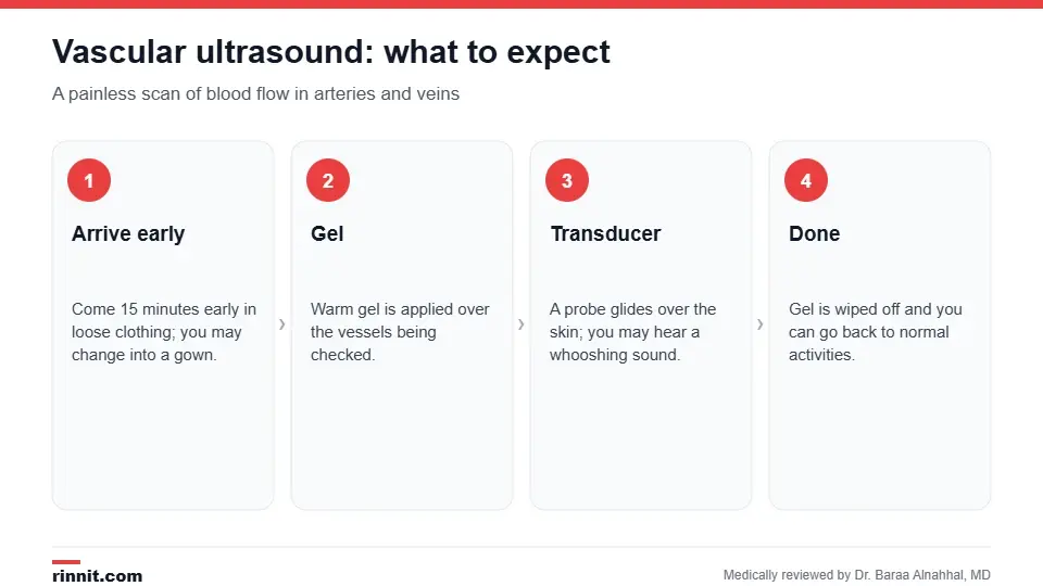
A vascular ultrasound is painless and has no side effects.
Why is it done?

Conditions found with vascular ultrasound.
Deep vein thrombosis (DVT) and other blood clots.
Peripheral artery disease and atherosclerosis.
Carotid artery disease, checked with a carotid ultrasound.
Aneurysms, such as an extracranial carotid artery aneurysm.
Checking whether you're a candidate for angioplasty, and blood flow after bypass surgery or an organ transplant. Sources: [1]
It may be ordered for leg pain or burning, pain in the buttocks, hips, thighs or calves when walking, muscle wasting or leg ulcers that won't heal. It often goes along with an ankle-brachial index test for leg arteries. Sources: [1]
How do I prepare?
Usually no special preparation is needed.
Arrive about 15 minutes early.
Wear comfortable, loose clothing and leave jewelry and valuables at home.
You may be asked to change into a gown.
If the scan includes vessels in the abdomen, you may be asked not to eat for several hours beforehand so gas doesn't block the view. Sources: [1]
What happens during the test?
You lie on an exam table.
The technologist applies a warm, water-based gel over the blood vessels being checked.
They press a handheld probe (transducer) against your skin and move it along the vessels. You may feel slight pressure and hear a pulsing or whooshing sound, which is blood flowing.
For veins, they may press on the vein or ask you to squeeze your calf to check for clots.
The gel is wiped off, and you can return to normal activities right away. Sources: [1]
Understanding your results

Vascular ultrasound is accurate for clots and artery disease.
A radiologist reviews the images and sends a report to your provider. The results show how fast or slowly blood flows, whether a clot or plaque is blocking a vessel, and whether vessels are narrowed or widened. Treatment depends on what's found, from blood thinners for a clot to lifestyle changes, medicine or procedures for narrowed arteries. Sources: [1, 2, 3]
Risks
Vascular ultrasound is safe. It uses no radiation and has no side effects. Sources: [1]
Contact your provider if symptoms such as pain, swelling or redness in the examined area get worse. Seek emergency care for sudden chest pain or shortness of breath, which can signal a blood clot in the lungs. Sources: [1]
Conclusion
A vascular ultrasound is a fast, safe way to see how blood moves through your arteries and veins. It's usually the first test for leg clots and artery disease, and it can guide treatment without radiation or needles.
Keep learning: read about DVT and peripheral artery disease.
Frequently asked questions
What does a vascular ultrasound check?
Blood flow through your arteries and veins, and blockages, clots or narrowing.
Is a vascular ultrasound painful?
No. You may feel slight pressure from the probe.
Do I need to prepare?
Usually not, though abdominal scans may require fasting.
Does it use radiation?
No. It uses sound waves.
Can it detect blood clots?
Yes. It's the main test for deep vein thrombosis.
What is a duplex ultrasound?
An ultrasound that shows both the vessel and the blood flowing through it using Doppler.
Why do I hear a whooshing sound?
That's the sound of blood flowing, picked up by the Doppler probe.
How accurate is it?
Very accurate; about 90% sensitive for DVT and 88% for significant leg artery narrowing.
Who performs the test?
An ultrasound technologist; a radiologist reads the images.
Can I go back to normal activities afterward?
Yes, right away.
Related Reading
Vein Mapping: Ultrasound Before Bypass Surgery or Dialysis Access explains the ultrasound that maps blood vessels before surgery or dialysis access.
Deep Vein Thrombosis (DVT): Symptoms, Causes, Diagnosis & Treatment — Complete Guide blood clots a vascular ultrasound finds.
Peripheral Artery Disease (PAD): Symptoms, Causes, Diagnosis & Treatment Options narrowed leg arteries seen on duplex scans.
Carotid Ultrasound: Why It's Done, What to Expect and What Results Mean a vascular ultrasound of the neck arteries.
Chronic Venous Insufficiency (CVI): What It Is, Why It Happens, and How It Gets Treated leaky leg veins checked with ultrasound.
What Are Varicose Veins? A Plain-Language Guide to the Bulging, Twisted Veins That Most Often Show Up on the Legs enlarged veins assessed before treatment.
Venous Extremity Ultrasound: Checking Arm and Leg Veins for Blood Clots explains the ultrasound that checks arm and leg veins for blood clots.
References
Cleveland Clinic. What is a Vascular Ultrasound? Cleveland Clinic. Last updated July 25, 2022. https://my.clevelandclinic.org/health/diagnostics/17606-vascular-ultrasound
Hercz D, Mechanic OJ, Varella M, et al. Ultrasound performed by emergency physicians for deep vein thrombosis: a systematic review. West J Emerg Med. 2024;25(2):282-290. PMID 38596931. https://doi.org/10.5811/westjem.18125
Collins R, Burch J, Cranny G, et al. Duplex ultrasonography, magnetic resonance angiography, and computed tomography angiography for diagnosis and assessment of symptomatic, lower limb peripheral arterial disease: systematic review. BMJ. 2007;334(7606):1257. PMID 17548364. https://doi.org/10.1136/bmj.39217.473275.55
Medical disclaimer
This article is for general education only and does not replace advice from a qualified healthcare professional. Talk to your doctor about your own diagnosis and treatment, and seek emergency care right away for severe or sudden symptoms.



Comments