Doppler Ultrasound: How It Works, Types, Uses and Results

Updated: 5 days ago
Written and medically reviewed by Dr. Baraa Alnahhal, MD · Last reviewed: November 2026 · How we review
Quick Answer: What is a Doppler ultrasound?
A Doppler ultrasound is a painless imaging test that uses sound waves to show how blood moves through your arteries and veins. It measures the speed and direction of blood flow, so doctors can find blood clots, narrowed or blocked vessels and other circulation problems in the legs, arms, belly, neck and heart. It needs no needles, dye or radiation. [1]
What the research says
Doppler-based scans find most leg clots above the knee. A meta-analysis of 100 study groups found that duplex ultrasound detected 96.5% of proximal deep vein thromboses (DVT) and 71.2% of clots below the knee, with 94.0% specificity. (Goodacre et al., BMC Medical Imaging, 2005) [2]
Umbilical artery Doppler is linked to fewer baby deaths in high-risk pregnancies. In 16 trials with 10,225 babies, Doppler monitoring was linked to fewer perinatal deaths (1.2% vs 1.7%; risk ratio 0.71), plus fewer inductions of labor and cesarean births. (Alfirevic et al., Cochrane Database of Systematic Reviews, 2017) [3]
Key takeaways
A Doppler ultrasound shows how fast blood flows and in which direction. [1]
It helps diagnose blood clots, narrowed arteries, vein problems and vessel injuries. [1]
The test takes 30 to 60 minutes, is painless and is safe in pregnancy. [1]
You may need to fast or avoid nicotine for at least two hours before some scans. [1]
Results can take about a week and may lead to more tests. [1]
How does a Doppler ultrasound work?
A Doppler ultrasound works by bouncing sound waves off moving red blood cells. A probe sends sound waves into your body, and the waves echo back from blood flowing through your vessels. A computer compares the pitch of the waves going in with the pitch of the echoes coming back. That change in pitch tells your care team how fast your blood is moving and which way it is going. [1]

The test can show: [1]
How fast or slow your blood is moving
Whether something is blocking blood flow
Blood flowing the wrong way or pooling inside a vessel
The method is named after Christian Doppler, a 19th-century physicist who described how sound waves change when they reflect off moving objects. This is called the Doppler effect. [1]
Types of Doppler ultrasound
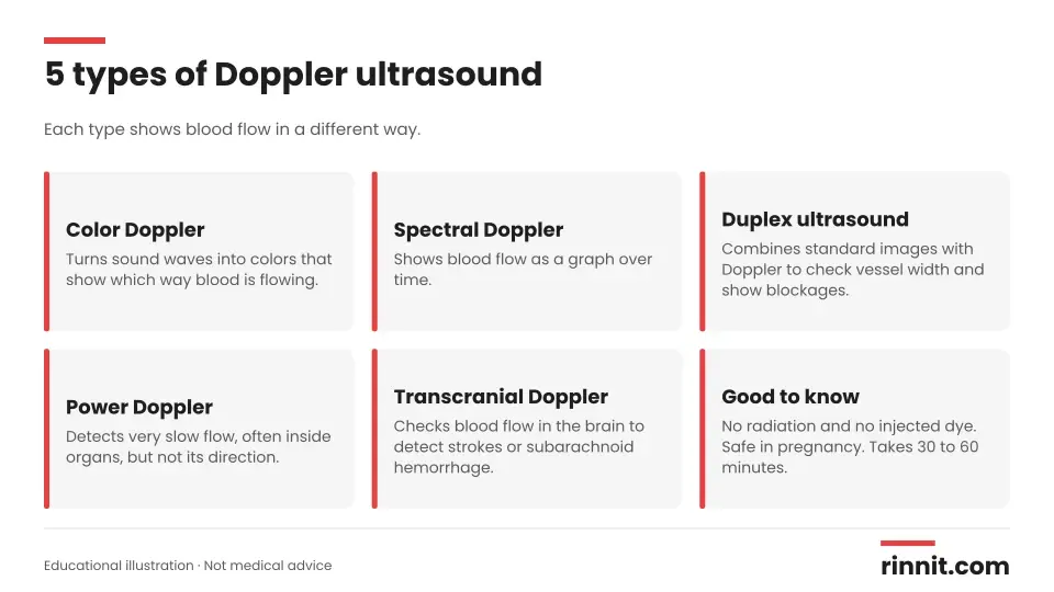
A standard ultrasound makes pictures but can't show blood flow. Doppler techniques add that information in different ways: [1]
Type | What it shows |
Color Doppler | Colors that show the direction of blood flow |
Spectral Doppler | A graph of blood flow over time |
Duplex ultrasound | Standard images plus Doppler, to check vessel width and show blockages |
Power Doppler | Very slow flow, often inside organs, without showing direction |
Transcranial Doppler | Blood flow in the brain, to detect strokes or subarachnoid hemorrhage |
Sources: [1]
Why would you need a Doppler ultrasound?
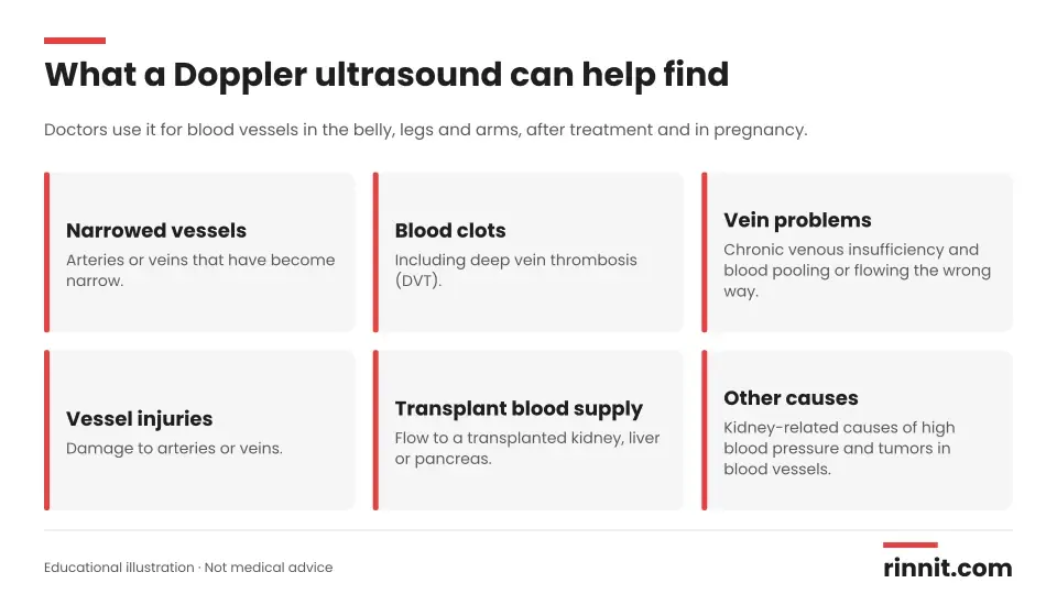
Your doctor may order a Doppler ultrasound to diagnose problems with blood vessels in your belly, legs or arms. It is also used to check blood flow after surgery or certain treatments, and to check blood flow between a pregnant person and the developing baby. [1]
Conditions a Doppler ultrasound can help diagnose include: [1]
Narrowed arteries or veins
Blood clots, including deep vein thrombosis (DVT)
Blood vessel injuries
Chronic venous insufficiency (CVI)
Problems with blood supply to a transplanted kidney, liver or pancreas
Kidney-related (renal vascular) causes of high blood pressure
Tumors in blood vessels
How do you prepare for a Doppler ultrasound?
Preparation depends on the type of scan and why you need it. You may be asked to: [1]
Fast (not eat or drink) for a set number of hours before the test
Stop smoking and avoid nicotine products for at least two hours before the test, because nicotine narrows blood vessels and can affect results
Ask your care team which instructions apply to your scan.
What happens during a Doppler ultrasound?
A sonographer, a specialist trained in ultrasound imaging, does the test. It usually takes 30 to 60 minutes. You may lie on your back or side, or sit up, depending on the area being checked. [1]
The sonographer puts a small amount of gel on your skin to help the probe glide and the sound waves travel.
The probe (transducer) sends painless, high-frequency sound waves through your skin. You won't hear them.
The sound waves reflect off moving blood cells, which changes their pitch. You may hear a whooshing sound from the machine.
The probe picks up the changes, and the machine turns them into images or graphs.
At the end, the sonographer wipes off the gel.
Sources: [1]
Is a Doppler ultrasound safe?
Yes. A Doppler ultrasound is a noninvasive, low-risk test. It doesn't use injected contrast dye like an angiogram, and it doesn't use radiation like X-rays or CT scans. It isn't painful, and it is safe enough to use during pregnancy. [1]
Results and follow-up
A radiologist (a doctor who specializes in medical imaging) or your own provider reviews the images. Results may take about a week. Depending on what the scan shows, you may need more tests, and the type of test will depend on the condition found. [1]
Questions worth asking your provider before the test: [1]
What type of ultrasound do I need?
How should I prepare?
When will I get my results?
Will I need other tests?
Conclusion
A Doppler ultrasound is a safe, painless way to see how blood moves through your heart and blood vessels. It helps your care team find clots, blockages and other circulation problems, and check whether treatment is working. [1]
If your doctor orders one, ask which type you need and whether you should fast or avoid nicotine first. If you have sudden leg swelling and pain, chest pain or shortness of breath, seek medical care right away rather than waiting for a scheduled test.
Keep learning: Read our guide to peripheral artery disease or learn about varicose veins. For more background, see the Doppler ultrasound overview from MedlinePlus.
FAQ
What is a Doppler ultrasound?
It is a painless imaging test that uses sound waves to show the speed and direction of blood flow in your arteries and veins. [1]
What is a Doppler ultrasound used for?
It helps diagnose blood clots, narrowed or blocked vessels, vein problems, vessel injuries and blood-supply problems in transplanted organs. [1]
Can a Doppler ultrasound detect blood clots?
Yes. It is commonly used to find blood clots, including deep vein thrombosis (DVT) in the legs. [1]
How long does a Doppler ultrasound take?
Most Doppler ultrasounds take about 30 to 60 minutes. [1]
Does a Doppler ultrasound hurt?
No. The probe glides over gel on your skin, and the sound waves are painless and can't be heard. [1]
How do I prepare for a Doppler ultrasound?
You may need to fast for a few hours and avoid nicotine for at least two hours, depending on the scan. [1]
Is a Doppler ultrasound safe during pregnancy?
Yes. It uses no radiation or contrast dye and is safe enough to use during pregnancy. [1]
What is the difference between a Doppler and a regular ultrasound?
A regular ultrasound makes images of organs and tissues, while a Doppler ultrasound also shows how blood is flowing. [1]
What is a duplex ultrasound?
A duplex ultrasound combines standard ultrasound images with Doppler to check vessel width and show blockages. [1]
How long do Doppler ultrasound results take?
A radiologist or your provider reviews the scan, and results may take about a week. [1]
Related Reading
Testicular Ultrasound: Checking Lumps, Pain and Swelling explains the ultrasound used to check scrotal lumps, pain and swelling.
Vein Mapping: Ultrasound Before Bypass Surgery or Dialysis Access explains the ultrasound that maps blood vessels before surgery or dialysis access.
Peripheral Artery Disease (PAD): Symptoms, Causes, Diagnosis & Treatment Options explains a circulation problem that a Doppler ultrasound helps diagnose.
Carotid Artery Disease: The Silent Stroke Risk in Your Neck, Explained covers narrowed neck arteries, often checked with a Doppler (duplex) scan.
What Are Varicose Veins? A Plain-Language Guide to the Bulging, Twisted Veins That Most Often Show Up on the Legs explains vein problems that a Doppler ultrasound can assess.
Narrowed Kidney Arteries: What Renal Artery Stenosis Is, Why It Happens, and How It Is Treated covers a renal vascular cause of high blood pressure that a Doppler scan can help find.
Point-of-Care Ultrasound (POCUS): What It Is, Uses and How It Differs From a Standard Scan explains the bedside ultrasound clinicians use for quick, focused answers.
Transcranial Doppler (TCD): Ultrasound of Blood Flow in the Brain explains the ultrasound test that measures blood flow in the brain.
References
Cleveland Clinic. Doppler Ultrasound: What Is It, Purpose and Procedure Details. Cleveland Clinic. Last updated April 12, 2022. https://my.clevelandclinic.org/health/diagnostics/22715-doppler-ultrasound
Goodacre S, Sampson F, Thomas S, et al. Systematic review and meta-analysis of the diagnostic accuracy of ultrasonography for deep vein thrombosis. BMC Med Imaging. 2005;5:6. PMID 16202135. https://doi.org/10.1186/1471-2342-5-6
Alfirevic Z, Stampalija T, Dowswell T. Fetal and umbilical Doppler ultrasound in high-risk pregnancies. Cochrane Database Syst Rev. 2017;6:CD007529. PMID 28613398. https://doi.org/10.1002/14651858.CD007529.pub4
Medical disclaimer
This article is for general education only and does not replace advice from a qualified healthcare professional. Talk to your doctor about your own diagnosis and treatment, and seek emergency care right away for severe or sudden symptoms.



Comments