Pathology Report: How to Read Margins, Grade, Stage and More

Updated: 3 days ago
Written and medically reviewed by Dr. Baraa Alnahhal, MD · Last reviewed: November 2026 · How we review
Quick Answer
A pathology report is a document written by a pathologist, a doctor who examines tissue and fluid samples under a microscope, describing what they found and giving a diagnosis. For cancer, it includes the tumor type, grade, stage, margins and lymph node findings. Results take a week or longer. Ask your provider about any terms you don't understand.
What the research says
A second look can change the diagnosis: among 2,786 blood-cancer pathology cases referred to an academic center for a second opinion, 9.4% had a different diagnosis and 5.9% had a major change that could affect treatment (Hamnvag HM et al., Am J Clin Pathol, 2025) [2]
Digital slides can speed up expert review: when head and neck pathology consultations were reviewed digitally and later rechecked on glass slides, there were no major discrepancies, and the digital system cut the median turnaround time by 97% (Liu BL et al., Am J Surg Pathol, 2024) [3]
Key takeaways
Written by a pathologist after examining a sample under a microscope.
Samples can be tissue, blood, urine, stool or other fluids.
Cancer reports describe type, grade, stage, margins and lymph nodes.
Molecular results can help choose targeted treatments.
Results often take a week or longer; your provider will explain them.
What is a pathology report?
Pathology is the study of disease in cells and tissues. A pathologist compares your sample with what normal cells and tissue should look like and writes a report with a diagnosis or recommendations. It helps diagnose, monitor or screen for disease, plan treatment, estimate outlook and, with genetic testing, assess risk. Sources: [1]
Pathology reports come from many tests, including a biopsy, cytology, urinalysis, a throat culture, surgery such as a lumpectomy, colonoscopy and endoscopy. Sources: [1]
How a sample becomes a report

Results usually take a week or longer.
A sample is taken by biopsy, surgery or fluid collection.
It's preserved, often in formalin, and processed into thin slides.
The pathologist examines it, often with stains that highlight cell parts or germs.
A report is sent to your provider. Sources: [1]
Sections of a pathology report
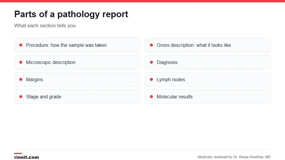
Each section answers a different question.
Section | What it tells you |
Procedure | How the sample was obtained |
Gross description | What the sample looks like without a microscope: size, weight, color |
Microscopic description | What the cells and tissue look like under magnification |
Margins | Whether cancer cells reached the edges of removed tissue |
Stage and grade | How far cancer has spread and how abnormal the cells look |
Lymph nodes | Whether cancer cells were found in nearby lymph nodes |
Molecular results | Gene changes in cancer cells that may guide treatment |
Pathogens | Bacteria, viruses, parasites or fungi, often in stool or fluid reports |
Diagnosis | The pathologist's summary |
Recommendations | Suggested follow-up |
Sources: [1]
Key terms to know

Negative margins and nodes are usually good news.
Negative (clear) margins: no cancer cells at the edges, suggesting all the cancer was removed.
Positive margins: cancer cells at the edge, so some cancer may remain.
Positive lymph nodes: cancer cells found in the nodes, as in a sentinel node biopsy or lymph node biopsy.
Grade: how different cancer cells look from normal; prostate cancer uses the Gleason score.
Stage: how far cancer has spread; see cancer staging and grading. Sources: [1]
How long does it take?
Results usually take a week or longer, though some come back sooner. Extra stains, special tests or a second opinion can add time. A delay doesn't mean anything about your result. Sources: [1, 2]
Reading your report
Pathology reports are written for doctors, so they can be hard to follow. Your provider will explain the results and next steps. Write down questions, ask what each term means for you, and ask whether a second opinion would be helpful for a complex diagnosis. Sources: [1, 2, 3]
Conclusion
A pathology report is often the key document behind a diagnosis, especially for cancer. Knowing its main sections, such as diagnosis, grade, stage, margins and lymph nodes, can help you have a clearer conversation with your care team.
Keep learning: read about biopsy and cancer staging and grading.
Frequently asked questions
What is a pathology report?
A pathologist's description and diagnosis of a tissue or fluid sample.
Who writes it?
A pathologist.
How long does it take?
Usually a week or longer.
What does negative margins mean?
No cancer cells were found at the edges of the removed tissue.
What does positive lymph nodes mean?
Cancer cells were found in the lymph nodes.
What's the difference between grade and stage?
Grade describes how abnormal cells look; stage describes how far cancer has spread.
What are molecular results?
Tests for gene changes that can guide treatment.
Does a delay mean bad news?
No. Delays don't indicate the result.
Can I get a second opinion?
Yes. Ask your provider about having the slides reviewed.
Who explains my report?
Your healthcare provider.
Related Reading
Immunostaining: How Labeled Antibodies Help Diagnose Cancer and Other Diseases explains the antibody staining methods pathologists use to pinpoint a diagnosis.
Biopsy: What It Is, Types, Preparation, Risks and Results how tissue samples are collected.
Cytology (Cytopathology): Types, Why It's Done, What Happens and What Results Mean the study of individual cells.
Cancer Staging and Grading: What They Are, Why They Matter, Tests and Results how cancer is staged and graded.
Sentinel Node Biopsy: How It Works, Recovery, Risks and Results a lymph node test often reported by pathology.
Gleason Score: How Prostate Cancer Is Graded and What Your Score Means the grading system for prostate cancer.
FISH Test: How Fluorescence In Situ Hybridization Finds Gene Changes explains the glowing-probe lab test that finds gene and chromosome changes.
References
Cleveland Clinic. Pathology Report: What It Is, How to Read It & Types. Cleveland Clinic. Last updated June 7, 2024. https://my.clevelandclinic.org/health/diagnostics/pathology-report
Hamnvag HM, Van Norman S, Chen Y, et al. Secondary review of extramural hematopathology cases for patients referred to an academic center: the increasing importance of subspecialized hematopathology practice. Am J Clin Pathol. 2025;164(5):800-805. PMID 40994294. https://doi.org/10.1093/ajcp/aqaf105
Liu BL, Haghighi M, Westra WH. Digital pathology is a fast and effective platform for providing head and neck pathology consultations. Am J Surg Pathol. 2024;48(8):985-990. PMID 38712588. https://doi.org/10.1097/PAS.0000000000002239
Medical disclaimer
This article is for general education only and does not replace advice from a qualified healthcare professional. Talk to your doctor about your own diagnosis and treatment, and seek emergency care right away for severe or sudden symptoms.



Comments