Immunostaining: How Labeled Antibodies Help Diagnose Cancer and Other Diseases

Updated: 2 days ago
Written and medically reviewed by Dr. Baraa Alnahhal, MD · Last reviewed: November 2026 · How we review
Quick Answer
Immunostaining is a lab method pathologists use to find a specific protein in a sample of tissue, blood or bone marrow. It uses antibodies that stick to their target and carry a label that makes it visible. It helps diagnose cancers, infections, blood disorders and autoimmune diseases. Results can take from a few hours to about 10 days.
What the research says
Immunostaining helps trace a cancer's origin: in carcinoma of unknown primary site, immunohistochemical staining predicted a single site of origin in about 35% to 55% of patients, and when it did, it agreed with gene expression profiling in 77% of cases (Hainsworth JD et al., Virchows Archiv, 2014) [2]
Flow cytometry is key for blood cancers: a review in Blood describes how flow cytometric immunophenotyping is used within the World Health Organization's multiparameter approach to diagnose and classify blood cancers and to guide further testing (Craig FE et al., Blood, 2008) [3]
Key takeaways
Immunostaining uses labeled antibodies to find specific proteins.
Immunohistochemistry on tissue slides is the most common type.
Flow cytometry uses lasers to sort and count labeled cells.
It helps diagnose cancer, infections, blood disorders and autoimmune disease.
Results can take a few hours to about 10 days.
What is immunostaining?
Cells carry many different proteins. Some, called antigens, are typical of a certain cell type, infection or cancer. In immunostaining, the lab adds antibodies designed to bind a chosen antigen. A label attached to the antibody then shows where, and how much, of that protein is present. A pathologist reads the result and reports it in a pathology report. Sources: [1]
Types of immunostaining
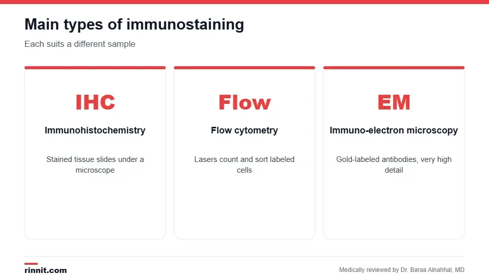
Each method suits a different kind of sample and question.
Type | How it works | Common uses |
Immunohistochemistry (IHC) | Stains a thin tissue slice on a glass slide, viewed under a microscope | Cancer diagnosis and typing, some neurological and blood disorders |
Flow cytometry | Lasers count and sort labeled cells one by one | Leukemia, lymphoma, infections, autoimmune disease |
Immuno-electron microscopy | Gold-labeled antibodies seen under an electron microscope | Viral infections and research |
How it works
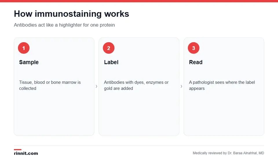
Antibodies work like a highlighter for one protein.
A provider collects a sample, such as a biopsy, blood or a bone marrow biopsy.
In the lab, antibodies with labels are added to the sample.
The labels make the target protein visible under a microscope or on a flow cytometer.
A pathologist interprets the pattern and sends a report to your provider. Sources: [1]
Why it's done

Immunostaining adds precision to a diagnosis.
Diagnose cancer and tell apart cancer types.
Help find where a cancer started.
Identify infections, including some viruses.
Diagnose and classify leukemia and lymphoma.
Help guide treatment by showing markers that targeted therapies act on. Sources: [1, 2, 3]
Preparation, timing and results
You usually don't need to do anything special; your provider will explain how the sample will be collected. Some immunostaining methods take a few hours, while others take up to 10 days. A pathologist analyzes the results and your provider explains them. If a disease is found, a team of doctors may plan treatment together. Sources: [1]
Conclusion
Immunostaining lets pathologists see specific proteins in your cells. It's a behind-the-scenes test that makes many diagnoses, especially cancer diagnoses, more precise.
Keep learning: read about immunohistochemistry and the pathology report.
Frequently asked questions
What is immunostaining?
A lab method that uses labeled antibodies to find specific proteins in a sample.
What samples are used?
Tissue, blood or bone marrow.
What is the most common type?
Immunohistochemistry on tissue slides.
Is immunostaining the same as immunohistochemistry?
Immunohistochemistry is one type of immunostaining.
What can it diagnose?
Cancers, infections, blood disorders and some autoimmune conditions.
Do I need to prepare?
Usually not, apart from instructions for the sample collection.
How long does it take?
From a few hours to about 10 days, depending on the method.
Who reads the results?
A pathologist.
Can it help choose cancer treatment?
Yes. It can show markers that some targeted treatments act on.
Is it painful?
The test itself is done in a lab; only the sample collection may be uncomfortable.
Related Reading
Immunohistochemistry (IHC): What It Is, How It Works and What Results Mean the most common type of immunostaining.
Flow Cytometry: What It Is, Why It's Done, How It Works and Results immunostaining for cells in fluid samples.
Pathology Report: How to Read Margins, Grade, Stage and More where immunostaining results appear.
Biopsy: What It Is, Types, Preparation, Risks and Results how tissue samples are collected.
Immunophenotyping: How It Identifies Blood Cells to Diagnose Leukemia and Lymphoma immunostaining used to classify leukemia and lymphoma.
References
Cleveland Clinic. What Is Immunostaining? Cleveland Clinic. Last updated June 19, 2023. https://my.clevelandclinic.org/health/diagnostics/25080-immunostaining
Hainsworth JD, Greco FA. Gene expression profiling in patients with carcinoma of unknown primary site: from translational research to standard of care. Virchows Archiv. 2014;464(4):393-402. PMID 24487792. https://doi.org/10.1007/s00428-014-1545-2
Craig FE, Foon KA. Flow cytometric immunophenotyping for hematologic neoplasms. Blood. 2008;111(8):3941-3967. PMID 18198345. https://doi.org/10.1182/blood-2007-11-120535
Medical disclaimer
This article is for general education only and does not replace advice from a qualified healthcare professional. Talk to your doctor about your own diagnosis and treatment, and seek emergency care right away for severe or sudden symptoms.



Comments