Immunophenotyping: How It Identifies Blood Cells to Diagnose Leukemia and Lymphoma

Updated: 2 days ago
Written and medically reviewed by Dr. Baraa Alnahhal, MD · Last reviewed: November 2026 · How we review
Quick Answer
Immunophenotyping is a lab test that identifies the types of white blood cells in a sample by the markers, called antigens, on their surface. It's most often done with flow cytometry on blood or bone marrow. Providers use it mainly to diagnose and classify leukemia and lymphoma and to track how well treatment is working.
What the research says
Flow cytometry is central to diagnosing blood cancers: a review in Blood describes how flow cytometric immunophenotyping, as part of the World Health Organization's multiparameter approach, is used to diagnose and classify blood cancers and guide other testing, following international consensus indications (Craig FE et al., Blood, 2008) [2]
Clearing leftover leukemia cells predicts better outcomes: a meta-analysis of 39 studies with 13,637 patients found that reaching minimal residual disease (MRD) negativity, often measured with flow cytometry, was linked to about a 77% lower risk of relapse or death in children and 72% in adults with acute lymphoblastic leukemia (Berry DA et al., JAMA Oncol, 2017) [3]
Key takeaways
Immunophenotyping identifies cells by the markers on their surface.
It's most often done with flow cytometry.
Samples are usually blood or bone marrow.
It helps diagnose and classify leukemia and lymphoma.
It can also detect small numbers of leftover cancer cells after treatment.
What is immunophenotyping?
Every type of white blood cell carries a typical set of surface proteins, often named with CD (cluster of differentiation) numbers. Cancer cells often show an unusual pattern. In immunophenotyping, the lab adds antibodies that stick to specific markers, then measures which cells light up. This shows what kinds of cells are present and whether a group of abnormal cells all came from the same source. Sources: [1, 2]
How it works

Flow cytometry reads thousands of cells per second.
The sample is mixed with antibodies tagged with fluorescent dyes.
In flow cytometry, cells flow one by one past a laser, and detectors record which dyes each cell carries.
A specialist reads the pattern of markers to identify cell types. Sources: [1, 2]
In immunohistochemistry, the antibodies are applied to a thin slice of tissue, such as a lymph node biopsy, and viewed under a microscope. Sources: [1]
Samples used
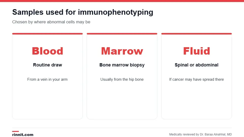
The sample depends on where abnormal cells are suspected.
Blood: a routine draw from a vein.
Bone marrow: collected with a bone marrow biopsy, usually from the hip bone.
Spinal fluid: collected with a lumbar puncture if cancer may have reached the nervous system.
Abdominal fluid or tissue: from a needle sample or biopsy. Sources: [1]
Why it's done
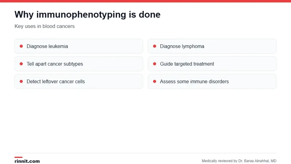
The test supports diagnosis, treatment choice and follow-up.
Diagnose leukemia and lymphoma after an abnormal blood count.
Tell apart subtypes, such as acute lymphocytic leukemia or chronic lymphocytic leukemia.
Help choose treatment, including targeted therapies aimed at specific markers.
Check for minimal residual disease during and after treatment.
Assess some immune system disorders. Sources: [1, 2, 3]
Understanding results
There isn't one standard procedure or normal value. A hematopathologist reports which cell types were found and whether there's an abnormal group of cells with a particular marker pattern. Results are read together with your blood count, bone marrow findings, genetic tests and symptoms. Sources: [1, 2]
Conclusion
Immunophenotyping helps providers see exactly which cells are present in a sample. It's a key tool for diagnosing and classifying blood cancers and for checking whether treatment has cleared the disease.
Keep learning: read about flow cytometry and leukemia.
Frequently asked questions
What does immunophenotyping test for?
The types of white blood cells in a sample, based on surface markers.
Is immunophenotyping the same as flow cytometry?
Flow cytometry is the most common method used to do it.
What samples are used?
Usually blood or bone marrow; sometimes spinal fluid, abdominal fluid or tissue.
Why would I need this test?
Most often to diagnose or classify leukemia or lymphoma.
Is the test painful?
A blood draw is quick; a bone marrow biopsy causes more discomfort.
What are CD markers?
Surface proteins labeled with numbers that identify cell types.
Can it detect cancer after treatment?
Yes. It can find minimal residual disease.
Is there a normal result?
There's no single normal value; a specialist interprets the pattern.
How long do results take?
It varies by lab and test, often several days.
What other tests are done with it?
Blood counts, bone marrow exams and genetic tests.
Related Reading
Immunostaining: How Labeled Antibodies Help Diagnose Cancer and Other Diseases explains the antibody staining methods pathologists use to pinpoint a diagnosis.
Flow Cytometry: What It Is, Why It's Done, How It Works and Results the main method used for immunophenotyping.
Immunohistochemistry (IHC): What It Is, How It Works and What Results Mean the tissue-based way to detect cell markers.
Understanding Leukemia: Types, Symptoms, and Modern Management the blood cancers this test helps classify.
Understanding Lymphoma: Types, Symptoms, and Modern Treatments another major use of immunophenotyping.
Bone Marrow Biopsy: Why It's Done, What to Expect, Pain and Recovery a common way to collect the sample.
References
Cleveland Clinic. What Is Immunophenotyping? Purpose, Testing & Results. Cleveland Clinic. Last updated July 1, 2024. https://my.clevelandclinic.org/health/diagnostics/immunophenotyping
Craig FE, Foon KA. Flow cytometric immunophenotyping for hematologic neoplasms. Blood. 2008;111(8):3941-3967. PMID 18198345. https://doi.org/10.1182/blood-2007-11-120535
Berry DA, Zhou S, Higley H, et al. Association of minimal residual disease with clinical outcome in pediatric and adult acute lymphoblastic leukemia: a meta-analysis. JAMA Oncol. 2017;3(7):e170580. PMID 28494052. https://doi.org/10.1001/jamaoncol.2017.0580
Medical disclaimer
This article is for general education only and does not replace advice from a qualified healthcare professional. Talk to your doctor about your own diagnosis and treatment, and seek emergency care right away for severe or sudden symptoms.



Comments