Immunohistochemistry (IHC): What It Is, How It Works and What Results Mean

Updated: 2 days ago
Written and medically reviewed by Dr. Baraa Alnahhal, MD · Last reviewed: November 2026 · How we review
Quick Answer
Immunohistochemistry (IHC) is a laboratory test performed on a biopsy or surgical tissue sample. Antibodies that recognize a specific protein are applied to thin slices of tissue, and a color shows where the protein is present. IHC helps identify a cancer's type and origin and decide whether targeted treatments such as hormone therapy or HER2-directed drugs will work.
What the research says
Some IHC markers are close to definitive: a 2026 meta-analysis of 23 studies found that STAT6 staining identifies solitary fibrous tumor with 98.7% sensitivity and 99.1% specificity, showing how a single well-validated antibody can settle a difficult diagnosis (Tayade V, Singh G, Annals of Diagnostic Pathology, 2026) [2]
IHC scores predict treatment response: across 13 studies and 7,206 patients with breast cancer, tumors scored HER2 3+ by IHC had a 55.1% complete response to HER2-targeted therapy versus 21.6% for HER2 2+/ISH-positive tumors (odds ratio 5.38) (Yucel KB et al., Cancer Treatment Reviews, 2026) [3]
Key takeaways
IHC is done on tissue you have already given; no extra procedure is needed.
Results are reported as positive or negative, often with a score such as 0, 1+, 2+ or 3+ and a percentage of stained cells.
It helps identify where a metastatic cancer started with roughly 70–90% accuracy.
ER, PR, HER2, PD-L1 and mismatch-repair proteins are among the markers that directly guide treatment.
Results typically take a few days to a week beyond the standard pathology report.
What is immunohistochemistry?
The first IHC stain was described in 1941, and the method is now routine in every pathology laboratory. It combines two ideas: the immune system's antibodies bind to one specific protein (antigen), and an enzyme such as horseradish peroxidase or alkaline phosphatase can be linked to that antibody to create a visible color reaction under the microscope. Sources: [1]
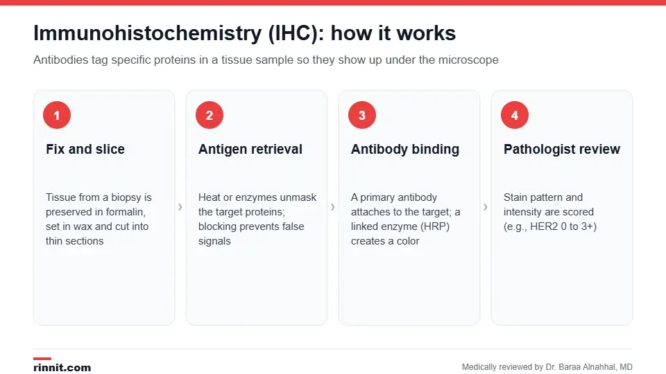
IHC adds targeted antibody stains to the standard tissue slides from a biopsy.
Why is IHC done?
Identify the cancer type when cells look similar under routine stains (for example lymphoma vs carcinoma vs melanoma).
Find the primary site of a metastasis using panels of tissue-specific markers.
Estimate prognosis, for example with proliferation markers such as Ki-67.
Guide treatment: estrogen and progesterone receptors and HER2 in breast cancer, androgen receptor in prostate cancer, PD-L1 for immunotherapy, mismatch-repair proteins for Lynch syndrome and immunotherapy.
Detect infections such as cytomegalovirus, herpes or Helicobacter pylori in tissue.
Confirm neurodegenerative disease by staining amyloid, tau or alpha-synuclein deposits. Sources: [1]
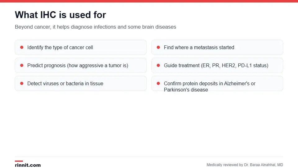
IHC answers questions a routine H&E stain cannot.
How is the test done?
Tissue is fixed in formalin, embedded in paraffin wax and cut into slices a few thousandths of a millimeter thick.
Heat or enzymes "unmask" the target protein (antigen retrieval), and a blocking step prevents antibodies sticking where they should not.
A primary antibody (monoclonal or polyclonal) is applied and binds the target.
A secondary antibody linked to an enzyme is added; a chromogen turns brown or red wherever the enzyme sits.
A counterstain outlines the cells, and a pathologist scores the pattern, intensity and percentage of positive cells. Sources: [1]
Nothing is required from you. IHC is performed on tissue already collected by biopsy or surgery, and it usually adds a few days to the final pathology report. Sources: [1]
Understanding your results
Marker | Where it is used | What a positive result means |
ER / PR | Breast cancer | Hormone therapy is likely to help |
HER2 (0, 1+, 2+, 3+) | Breast and gastric cancer | 3+ predicts strong response to HER2-targeted drugs; 2+ needs a confirmatory FISH test |
PD-L1 | Lung, bladder, head and neck cancers | Higher scores favor immunotherapy |
MLH1, MSH2, MSH6, PMS2 | Colorectal and endometrial cancer | Loss of a protein suggests mismatch-repair deficiency or Lynch syndrome |
Ki-67 | Many tumors | Higher percentage means faster-growing cells |
CD20, CD3, CD30 | Lymphoma | Defines the lymphoma subtype |
Scores are interpreted in context. In breast cancer, for instance, 13 studies covering 7,206 patients showed that an IHC score of HER2 3+ was associated with a 55.1% complete response to HER2-targeted therapy versus 21.6% in HER2 2+/ISH-positive tumors. Sources: [3]
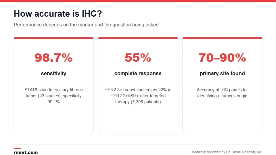
How accurate IHC is depends on the marker and the question asked.
How accurate is IHC?
Panels of markers identify the origin of a metastatic cancer in about 70–90% of cases. For some diagnoses a single stain is almost definitive: STAT6 identifies solitary fibrous tumor with 98.7% sensitivity and 99.1% specificity across 23 studies. Accuracy depends on tissue handling, antibody quality and the pathologist's interpretation, so results that do not fit the clinical picture may be repeated or supplemented with molecular tests. Sources: [1, 2]
Limitations
Poorly fixed or very small samples can give false-negative results.
Some proteins are present in both benign and malignant cells, so IHC is read together with the routine stain and the clinical history.
Equivocal scores (such as HER2 2+) need a confirmatory test like FISH.
IHC shows protein presence, not gene mutations; genetic sequencing may still be needed. Sources: [1]
Conclusion
Immunohistochemistry turns a slice of tissue into a map of the proteins that drive disease. For many people with cancer, it is the test that decides whether a targeted drug or hormone therapy is an option. Ask your care team which markers were tested on your sample and what each score means for your treatment plan.
Frequently asked questions
What is immunohistochemistry used for?
Mainly to classify cancers, find where a metastasis started, predict behavior and choose targeted treatments. It also detects infections and protein deposits in brain disease.
Do I need another biopsy for IHC?
Usually not. IHC is performed on the tissue already collected, as long as enough material remains.
How long does IHC take?
Typically a few days to a week after the initial pathology report, depending on how many stains are needed.
What does "HER2 3+" mean?
It means strong, complete staining in more than 10% of tumor cells, which predicts a good response to HER2-targeted therapy.
What does a negative IHC result mean?
The target protein was not detected. Depending on the marker, that may rule out a diagnosis or mean a particular drug is unlikely to help.
Is IHC the same as a genetic test?
No. IHC detects proteins in tissue. Genetic tests read DNA or RNA. They are often used together.
How reliable is IHC?
Very reliable for well-validated markers, with some stains exceeding 98% sensitivity and specificity. Accuracy depends on sample quality and interpretation.
Can IHC tell which organ a cancer came from?
Panels of markers identify the primary site in roughly 70–90% of metastatic cancers of unknown origin.
Is IHC used outside cancer?
Yes, to detect viruses and bacteria in tissue and to confirm Alzheimer's, Parkinson's and other neurodegenerative diseases, often at autopsy.
Who interprets the IHC result?
A pathologist scores the slides and includes the findings in your pathology report, which your treating doctor explains to you.
Related Reading
Immunostaining: How Labeled Antibodies Help Diagnose Cancer and Other Diseases explains the antibody staining methods pathologists use to pinpoint a diagnosis.
Immunophenotyping: How It Identifies Blood Cells to Diagnose Leukemia and Lymphoma explains the marker test used to diagnose and classify leukemia and lymphoma.
Biopsy: What It Is, Types, Preparation, Risks and Results how the tissue tested by IHC is collected.
Breast Biopsy: Types, What to Expect, Recovery and Results breast tissue sampling where ER, PR and HER2 staining is routine.
Lymphoma: Symptoms, Types, Causes, Diagnosis and Treatment a cancer whose subtypes are defined largely by IHC markers.
Hormone Receptor-Positive (HR+) Breast Cancer: Understanding Your Diagnosis what an ER/PR-positive IHC result means.
Navigating HER2-Negative Breast Cancer: Types, Treatments, and Outlook what a HER2-negative score means for treatment.
References
Cleveland Clinic. What Is Immunohistochemistry? Cleveland Clinic. Last updated June 20, 2023. https://my.clevelandclinic.org/health/diagnostics/25090-immunohistochemistry
Tayade V, Singh G. Diagnostic accuracy of nuclear STAT6 immunohistochemistry for solitary fibrous tumour: a systematic review and meta-analysis. Annals of Diagnostic Pathology. 2026;85:152703. PMID 42566929. https://doi.org/10.1016/j.anndiagpath.2026.152703
Yucel KB, Ilhan Y, Almuradova E, et al. Comparison of the pathological complete response between immunohistochemistry HER2 (3+) and HER2 (2+)/ISH+ early-stage breast cancer: a systematic review and meta-analysis. Cancer Treatment Reviews. 2026;143:103074. PMID 41512603. https://doi.org/10.1016/j.ctrv.2025.103074
Medical disclaimer
This article is for general education only and does not replace advice from a qualified healthcare professional. Talk to your doctor about your own diagnosis and treatment, and seek emergency care right away for severe or sudden symptoms.



Comments