20-Week Ultrasound (Anatomy Scan): What It Checks, How to Prepare and What Results Mean

Updated: 2 days ago
Written and medically reviewed by Dr. Baraa Alnahhal, MD · Last reviewed: November 2026 · How we review
Quick Answer: What is a 20-week ultrasound?
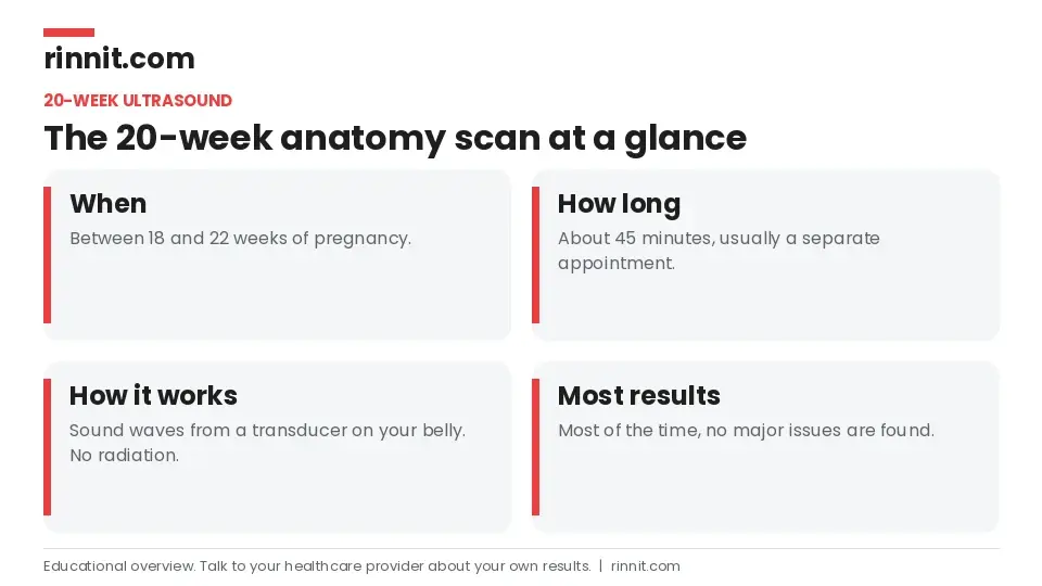
A 20-week ultrasound, also called an anatomy scan, is a prenatal ultrasound done between 18 and 22 weeks. It checks how the fetus is growing, looks at its organs and body parts, and can find signs of certain congenital conditions. The scan is safe, takes about 45 minutes, and most of the time no major problems are found. [1]
What the research says
The anatomy scan finds many, but not all, heart defects. A review of 7 population studies found the second-trimester scan detected 45.1% of congenital heart defects overall, but more than 85% of the most severe single-ventricle defects. Detection rose with how serious the defect was. (van Velzen et al., International Journal of Gynecology & Obstetrics, 2018) [2]
A suspected problem is not always a real one. In 526 pregnancies in France with a suspected malformation on second- or third-trimester ultrasound, 8.8% turned out to be false positives, most often for kidney and bowel findings. This is why a concerning result leads to expert follow-up rather than a final diagnosis. (Debost-Legrand et al., BMC Pregnancy and Childbirth, 2014) [3]
Key takeaways
The 20-week ultrasound happens between 18 and 22 weeks of pregnancy. [1]
A sonographer measures and photographs the baby's heart, brain, spine, kidneys, limbs, face and more. [1]
It also checks the placenta's position, the umbilical cord and the amount of amniotic fluid. [1]
There is little to prepare; the appointment takes about 45 minutes. [1]
The scan does not diagnose conditions. If something looks unusual, your provider orders more tests. [1]
In most cases you can learn the baby's sex, if you want to. [1]
What is the 20-week ultrasound?
The 20-week ultrasound is one of the main tools your pregnancy care provider uses to check that the fetus and the pregnancy are healthy. It helps them catch possible problems before birth, so you and your team can plan for any extra care your baby may need. [1]
What does the scan check?
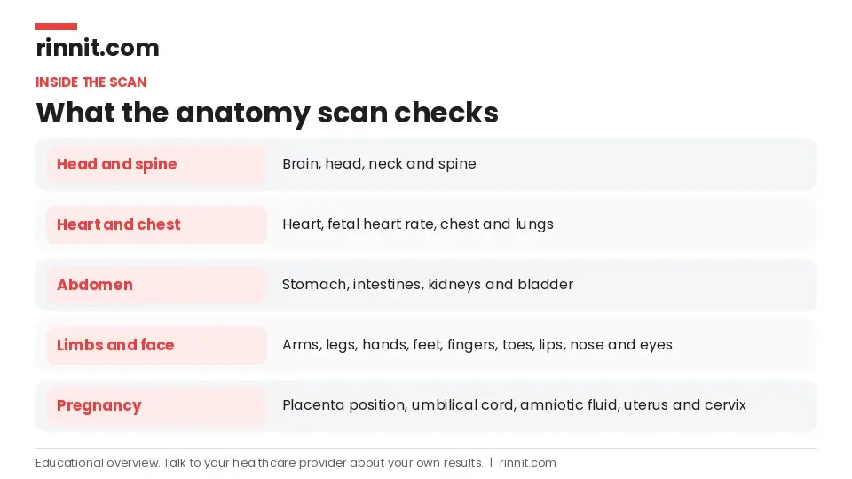
During your appointment, an ultrasound technician (sonographer) takes pictures and measurements of the fetal: [1]
Heart
Brain, head, neck and spine
Kidneys and bladder
Arms and legs
Hands, fingers, feet and toes
Lips, chin, nose, eyes and face
Chest and lungs
Stomach and intestines
They also record the fetal heart rate, check blood flow in the umbilical cord and where it attaches to the placenta, look at where the placenta sits, measure the amniotic fluid, and check your uterus, ovaries and cervix. [1]
How does a 20-week ultrasound work?
Ultrasound uses sound waves, not radiation, to make pictures of the inside of your body. The technician moves a handheld device called a transducer across your belly to get images of the fetus and takes measurements of its organs and body parts. [1]
How do you prepare?
There is not much to do. Plan for one of the longer prenatal ultrasound appointments, about 45 minutes, which is usually separate from your regular pregnancy visit. Some providers suggest eating beforehand, because it may make the fetus move more and help the technician get all the views they need. [1]
What happens during the scan?
You lie down on the exam table. [1]
The sonographer asks you to lower your pants slightly and lift your shirt, then puts ultrasound gel on your belly. [1]
They move the transducer over different spots, freezing the screen to capture images and measurements of specific organs. [1]
You may see them draw lines on the screen to measure the length of the limbs, head or belly. [1]
If the fetus is in a hard-to-measure position, you may be asked to move a little or have a sweet drink. [1]
When the scan is complete, they wipe off the gel. Many technicians send you home with a few pictures. [1]
The sonographer is often quiet while working, so do not be alarmed if they do not share results as they go. Your obstetrician or other provider goes over the results with you right after the scan or at a follow-up visit in the next few days. [1]
Will I find out the sex?
Usually, yes. Depending on the baby's position, the technician may be able to see the genitals. If you want it to be a surprise, tell them before the scan starts. [1]
Is the 20-week ultrasound safe?
Yes. The scan is not dangerous for you or the fetus. The main drawback is the chance of a false result. The benefits, finding conditions early so your care team can plan, outweigh this. [1]
What do the results mean?
The scan shows whether the fetus is growing at a healthy rate for its age. It also looks for signs of congenital conditions and structural problems in major organs such as the brain, heart and kidneys, checks the placenta's position and makes sure there is a safe amount of amniotic fluid. [1]
Most of the time, no major issues are found. The 20-week ultrasound does not diagnose anything on its own. If your provider sees something of concern, they will order more tests. [1]
What conditions can it help detect?
The scan does not find every congenital condition, but it can help detect several serious ones: [1]
Body area | Examples of conditions |
Brain and spine | Anencephaly, spina bifida |
Heart | Congenital heart abnormalities |
Face | Cleft lip |
Chromosomes | Signs of Down syndrome, trisomy 18 and trisomy 13 |
Abdomen and chest | Omphalocele, diaphragmatic hernia |
Kidneys | Renal agenesis (missing kidney) |
Skeleton | Skeletal dysplasia |
Sources: [1]
What happens if something looks unusual?
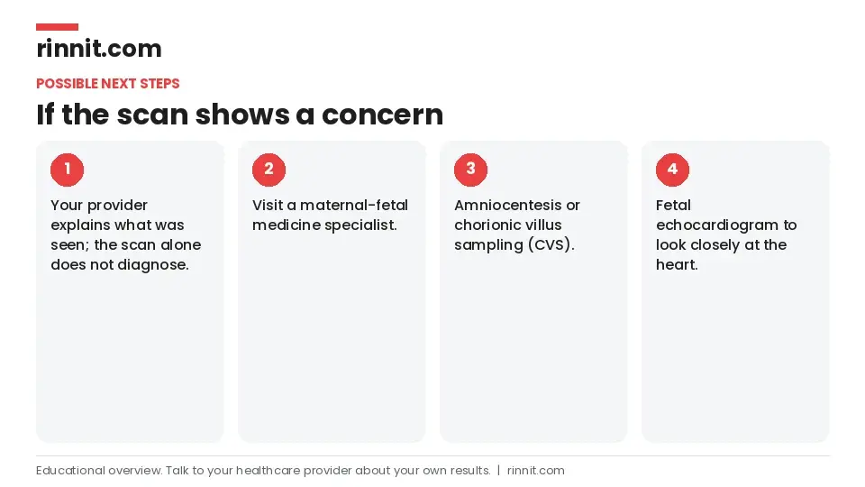
If your provider has concerns, they may recommend: [1]
A visit with a maternal-fetal medicine specialist, a doctor who cares for high-risk pregnancies
Talk with your provider early in pregnancy about noninvasive prenatal testing, because some screening tests can be done before the 20-week scan. [1]
Questions to ask at your appointment
Does everything look OK?
How big is my baby?
What are you looking at, and what measurements are you taking?
Will I know my results today?
What happens if something doesn't look right?
Is this test safe?
Is this the last ultrasound I'll have during pregnancy?
Can you tell if it's a boy or a girl?
Sources: [1]
Conclusion
The 20-week anatomy scan is a safe, detailed check of your baby's growth and organs at the halfway point of pregnancy. For most parents it is a reassuring experience. When it does show something unusual, it gives your care team time to arrange more testing and plan the right care after birth. [1]
It is normal to feel both excited and nervous. Bring your questions, and ask your provider to explain anything you see on the screen.
Keep learning: Read our guide to the fetal echocardiogram or our guide to congenital heart disease. For more trusted, doctor-reviewed health guides, visit Rinnit.com.
FAQ
What is a 20-week ultrasound?
It is a prenatal ultrasound at 18 to 22 weeks that checks the baby's growth, organs, placenta and amniotic fluid. [1]
Why is it called an anatomy scan?
Because the sonographer looks closely at the baby's anatomy, including the heart, brain, spine, kidneys, limbs and face. [1]
How long does the 20-week ultrasound take?
Plan for about 45 minutes. [1]
Do I need to prepare for the anatomy scan?
Not much. Some providers suggest eating beforehand so the baby moves more. [1]
Can the 20-week ultrasound tell the baby's sex?
Usually yes, depending on the baby's position. Tell the technician if you do not want to know. [1]
Is the 20-week ultrasound safe?
Yes. It uses sound waves and is not dangerous for you or the baby. [1]
What conditions can the anatomy scan detect?
It can help detect heart defects, spina bifida, anencephaly, cleft lip, signs of Down syndrome and some other conditions. [1]
Does the scan find every birth defect?
No. It does not find all congenital conditions, and it does not diagnose them on its own. [1]
When will I get my results?
Often right after the scan, or at a follow-up visit within a few days. [1]
What happens if the scan shows a problem?
Your provider may refer you to a maternal-fetal medicine specialist or suggest amniocentesis, CVS or a fetal echocardiogram. [1]
Related Reading
Fetal Biometry: Ultrasound Measurements of Baby's Growth Explained explains the ultrasound measurements used to track fetal growth.
Congenital Heart Disease (CHD): Types, Symptoms, Treatment & Survival — Complete Guide explains heart defects present at birth, one of the main things the anatomy scan looks for.
Down Syndrome: Symptoms, Causes, Types & Life Expectancy — Complete Guide covers Down syndrome, a chromosome condition the 20-week scan can show signs of.
Edwards Syndrome (Trisomy 18): Symptoms, Causes, Diagnosis, Treatment, Survival Rates and When to Seek Emergency Care — What You Need to Know covers trisomy 18, another chromosome condition the scan can help detect.
What Is Cleft Lip and Cleft Palate? Symptoms, Causes, Diagnosis and Treatment explains cleft lip and palate, which the scan may pick up on views of the face.
Anencephaly: What It Is, Why It Happens, and What Families Should Know describes a serious brain and skull condition the anatomy scan can detect.
What Is Omphalocele? Causes, Types, and Treatment explains an abdominal wall defect that can be seen on the scan.
Congenital Diaphragmatic Hernia (CDH): What It Is, Why It Happens, and How It's Treated covers a hole in the diaphragm that the 20-week ultrasound can help find.
High-Risk Pregnancy: Understanding Management, Factors, and Care explains the extra care that may follow if the scan finds a concern.
Fetoscopy: How It Treats Babies Before Birth, Procedure and Risks explains the camera procedure used to treat some conditions before birth.
References
Cleveland Clinic. 20 Week Ultrasound (Anatomy Scan): What To Expect. Cleveland Clinic. Last updated June 4, 2025. https://my.clevelandclinic.org/health/diagnostics/22644-20-week-ultrasound
van Velzen CL, Ket JCF, van de Ven PM, et al. Systematic review and meta-analysis of the performance of second-trimester screening for prenatal detection of congenital heart defects. Int J Gynaecol Obstet. 2018;140(2):137-145. PMID 29094357. https://doi.org/10.1002/ijgo.12373
Debost-Legrand A, Laurichesse-Delmas H, Francannet C, et al. False positive morphologic diagnoses at the anomaly scan: marginal or real problem, a population-based cohort study. BMC Pregnancy Childbirth. 2014;14:112. PMID 24655605. https://doi.org/10.1186/1471-2393-14-112
Medical disclaimer
This article is for general education only and does not replace advice from a qualified healthcare professional. Talk to your doctor about your own diagnosis and treatment, and seek emergency care right away for severe or sudden symptoms.



Comments