Fetal Echocardiogram: What It Checks, When It's Done and What Results Mean

Updated: 4 days ago
Written and medically reviewed by Dr. Baraa Alnahhal, MD · Last reviewed: November 2026 · How we review
Quick Answer: What is a fetal echocardiogram?
A fetal echocardiogram (fetal echo) is a detailed ultrasound of a baby's heart during pregnancy. It shows how the heart is forming, how it beats and how blood flows through it, so your care team can find congenital heart defects before birth and plan treatment for after delivery. It is usually done between 18 and 22 weeks. [1]
What the research says
Fetal echo is very specific and finds most defects in high-risk pregnancies. A meta-analysis of prenatal echocardiography found a pooled sensitivity of 68.5% and specificity of 99.8% for congenital heart disease; sensitivity rose to 85.1% in high-risk pregnancies versus 45.4% in low-risk ones. (Zhang et al., Medicine, 2015) [2]
Knowing before birth can save lives. Across eight studies of newborns with critical congenital heart disease, babies diagnosed before birth were less likely to die before their planned heart surgery than babies diagnosed after birth (pooled odds ratio 0.26). (Holland et al., Ultrasound in Obstetrics & Gynecology, 2015) [3]
Key takeaways
A fetal echo is a detailed ultrasound focused on the baby's heart and great vessels. [1]
It is usually done at 18 to 22 weeks, or as early as 14 to 16 weeks when a problem is suspected. [1]
Most scans are done through the belly; a transvaginal scan is less common. [1]
Pauses during the scan for measurements are routine and do not mean something is wrong. [1]
Unclear results usually lead to a repeat scan, not a diagnosis. [1]
What is a fetal echocardiogram?
A fetal echocardiogram uses very high-frequency sound waves to create a moving picture of the baby's heart, inside and out. It shows the heart beating and even how the valves inside it work. It can help diagnose heart defects, rule out suspected conditions and guide treatment planned for after birth. [1]
It works much like a regular pregnancy ultrasound, but it is more detailed and looks for specific heart problems. [1]
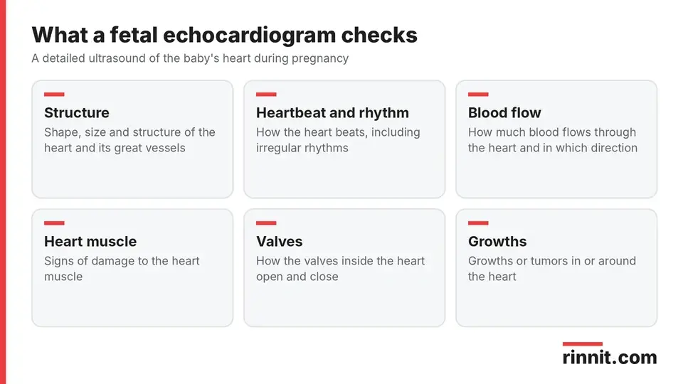
A fetal echo looks at the heart's structure, rhythm, blood flow and muscle.
A fetal echo looks at: [1]
The shape, size and structure of the heart and great vessels
How the heart beats
How much blood flows through the heart and in which direction
Muscle damage
Growths or tumors
Irregular heart rhythms
Why would a baby need a fetal echocardiogram?
Your provider may recommend a fetal echo if: [1]
At least one biological parent has a history of congenital heart defects
The fetus may have been exposed to an illness or substance that can affect development
You notice changes in how the fetus behaves during pregnancy
You already have a child with a congenital heart condition
The fetus has other congenital conditions
Your family has genetic diseases that can affect heart development
Bloodwork or amniocentesis results suggest a condition that may affect development
Other tests, such as a fetal electrocardiogram, show an irregular heart rhythm
When is a fetal echocardiogram done?
A fetal echo usually happens between 18 and 22 weeks of pregnancy. It can be done as early as 14 to 16 weeks, because some heart defects are visible that early, but early scans are mostly used when screening for an inherited condition or when a heart problem is already suspected. [1]
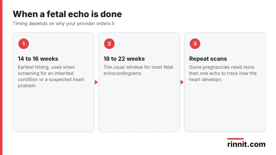
Most fetal echoes happen in the second trimester.
Some pregnancies need more than one fetal echo, so the care team can follow how the heart develops over time. That pattern can be a key clue to diagnosing certain conditions. [1]
What happens during a fetal echocardiogram?
There are two ways to do a fetal echo. The more common way is through the skin of your belly (abdominal); the less common way uses a small probe placed in the vagina (transvaginal). [1]

The abdominal approach is the most common.
Step | Abdominal fetal echo | Transvaginal fetal echo |
Getting ready | You may change into a gown and lie on your back on a reclining table | You may change into a gown or undress from the waist down under a drape |
The probe | Gel goes on your belly and the transducer moves across it | A small, wand-like transducer is placed in the vagina, like a tampon |
The scan | The tech views the fetus from all the needed angles | The tech gets a direct look at the fetus |
Sources: [1]
During either method, the technician may pause to take notes, make measurements and save images. This is a normal part of the scan and does not automatically mean they found a concern. [1]
What do fetal echocardiogram results mean?
After the scan, your healthcare provider shares the results and tells you whether everything looked normal or whether they saw anything concerning. [1]
If you have questions about what the scan shows, ask your provider. A recommendation for a fetal echo can bring up strong emotions, and your care team can help you understand what is happening and support you through the process. [1]
Conclusion
A fetal echocardiogram is a safe, detailed ultrasound that gives your care team a clear look at your baby's heart before birth. Most are done at 18 to 22 weeks through the belly, and many end with reassuring results.
If your provider recommends one, ask why, what they are looking for and whether you may need a repeat scan. Finding a heart problem early gives your team time to plan the right care for delivery and the first days of life.
Keep learning: Read our congenital heart disease guide or our guide to congenital heart defects in children. For more background, see the CDC page on congenital heart defects.
FAQ
What is a fetal echocardiogram?
It is a detailed ultrasound of a baby's heart during pregnancy that shows its structure, rhythm and blood flow.
Why would my baby need a fetal echo?
Common reasons include a family history of congenital heart defects, possible exposure to a harmful illness or substance, other birth differences or an irregular fetal heart rhythm.
When is a fetal echocardiogram done?
Usually between 18 and 22 weeks of pregnancy, and as early as 14 to 16 weeks when a problem is suspected.
How is a fetal echo different from a regular pregnancy ultrasound?
It works the same way but is more detailed and looks for specific heart problems.
Is a fetal echocardiogram done through the belly or the vagina?
Most are done through the belly; a transvaginal approach is used less often.
Does a fetal echocardiogram hurt?
No. It uses gel and a handheld probe, and it does not use radiation.
What can a fetal echo detect?
Problems with heart structure, the great vessels, heart rhythm, blood flow, heart muscle and growths.
What if the results are unclear?
Your provider may repeat the scan, because the baby's position or stage of development can limit the view.
Can I have more than one fetal echo?
Yes. Some pregnancies need several scans to track how the heart develops.
What happens if a problem is found?
Your provider explains the findings and plans next steps, which can include care right after birth.
Related Reading
Congenital Heart Disease (CHD): Types, Symptoms, Treatment & Survival — Complete Guide explains the heart conditions a fetal echo is designed to find.
Congenital Heart Defects in Children: Symptoms, Causes, and Treatment covers what happens after birth when a defect is found.
Tetralogy of Fallot: A Parent's Guide to the Heart Condition Babies Are Born With — Symptoms, Diagnosis, and Surgery describes one defect often diagnosed before birth.
Hypoplastic Left Heart Syndrome (HLHS): What It Is, Symptoms, Surgery Stages, and Lifelong Care explains a critical defect where planning before delivery matters.
Fetoscopy: How It Treats Babies Before Birth, Procedure and Risks explains the camera procedure used to treat some conditions before birth.
References
Cleveland Clinic. Fetal Echocardiogram: Procedure, Conditions & Treatment. Cleveland Clinic. Last updated June 19, 2026. https://my.clevelandclinic.org/health/diagnostics/fetal-echocardiogram
Zhang YF, Zeng XL, Zhao EF, et al. Diagnostic Value of Fetal Echocardiography for Congenital Heart Disease: A Systematic Review and Meta-Analysis. Medicine (Baltimore). 2015;94(42):e1759. PMID 26496297. https://doi.org/10.1097/MD.0000000000001759
Holland BJ, Myers JA, Woods CR. Prenatal diagnosis of critical congenital heart disease reduces risk of death from cardiovascular compromise prior to planned neonatal cardiac surgery: a meta-analysis. Ultrasound Obstet Gynecol. 2015;45(6):631-638. PMID 25904437. https://doi.org/10.1002/uog.14882
Medical disclaimer
This article is for general education only and does not replace advice from a qualified healthcare professional. Talk to your doctor about your own diagnosis and treatment, and seek emergency care right away for severe or sudden symptoms.



Comments