Fetoscopy: How It Treats Babies Before Birth, Procedure and Risks

Updated: 3 days ago
Written and medically reviewed by Dr. Baraa Alnahhal, MD · Last reviewed: November 2026 · How we review
Quick Answer
Fetoscopy is a procedure that uses a thin, lighted camera (fetoscope) passed into the uterus to see and treat a baby before birth. It's usually done after 18 weeks of pregnancy. Doctors use it to treat serious conditions such as twin-to-twin transfusion syndrome and amniotic band syndrome. Risks include waters breaking early, preterm birth and pregnancy loss.
What the research says
Laser fetoscopy improves twin survival: in a randomized trial of 142 pregnancies with severe twin-to-twin transfusion syndrome before 26 weeks, fetoscopic laser surgery led to at least one twin surviving to 28 days in 76% of pregnancies, compared with 56% after repeated fluid drainage, with fewer brain injuries (Senat MV et al., New England Journal of Medicine, 2004) [2]
Fetoscopic balloon treatment helps severe diaphragmatic hernia: in the TOTAL trial of babies with severe left-sided congenital diaphragmatic hernia, placing a balloon in the windpipe by fetoscopy at 27–29 weeks raised survival to hospital discharge to 36%, versus 14% with standard care, but increased early rupture of membranes and preterm birth (Deprest JA et al., New England Journal of Medicine, 2021) [3]
Key takeaways
A tiny camera inside the uterus to diagnose and treat a baby before birth.
Usually done after 18 weeks, through a small cut in the belly or, rarely, through the cervix.
Used for twin-to-twin transfusion syndrome, amniotic bands, diaphragmatic hernia, some spina bifida repairs and biopsies.
Less invasive than open fetal surgery; many people go home soon after.
Risks: early rupture of membranes, preterm labor and birth, pregnancy loss, infection and bleeding.
What is fetoscopy?
A fetoscope is a hollow fiber-optic tube with a camera. Once inside the amniotic sac, it sends images to a screen, and tiny instruments or a laser fiber can be passed through it. Because it needs only a small opening, fetoscopy is a form of minimally invasive fetal surgery. Sources: [1]
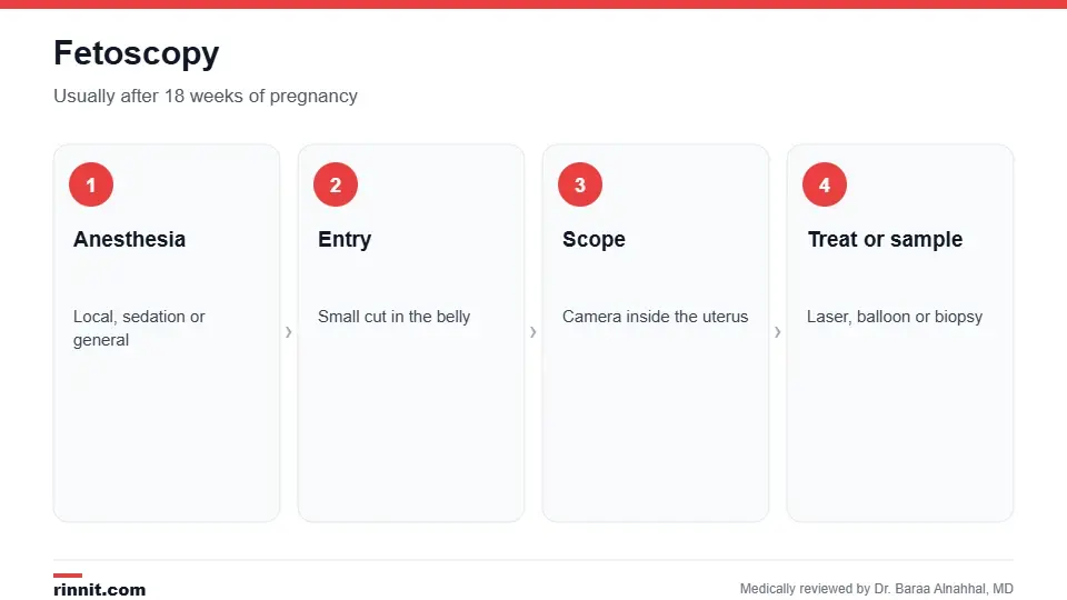
Fetoscopy is usually done after 18 weeks of pregnancy.
Why is it done?

Fetoscopy can treat several serious conditions before birth.
Twin-to-twin transfusion syndrome (TTTS): in identical twins sharing a placenta, a laser closes abnormal connecting blood vessels so blood is no longer unevenly shared.
Amniotic band syndrome: a laser cuts bands of tissue wrapped around the baby.
Congenital diaphragmatic hernia (CDH): a balloon placed in the windpipe (FETO) and removed weeks later helps the lungs grow.
Some spina bifida repairs and placental tumors.
Collecting tissue samples to diagnose rare conditions. Sources: [1]
Problems are usually found first on a 20-week anatomy scan or a fetal echocardiogram. Genetic testing, such as amniocentesis, may also be done before deciding on treatment. Sources: [1]
How do I prepare?
Preparation depends on the condition being treated and whether the scope goes through the belly or the cervix. Your fetal medicine team will explain fasting, medicines and what to bring, and discuss risks, benefits and delivery planning. Sources: [1]
What happens during the procedure?
You receive local anesthesia, sedation or sometimes general anesthesia; medicine may also be given to keep the baby still.
Using ultrasound guidance, the surgeon makes a small cut in the belly, or inserts the scope through the cervix with a speculum.
The fetoscope is passed into the uterus and amniotic sac, and images appear on a screen.
Instruments, a laser fiber or a balloon are passed through the scope to treat the baby or collect tissue. Sources: [1]
Many people return to normal activities quickly, though you'll be monitored closely afterward and asked to take care around the entry site. Sources: [1]
Risks

Your team will weigh these risks against the benefits.
Waters breaking early (preterm premature rupture of membranes).
Preterm labor and preterm birth.
Pregnancy loss.
Infection.
Bleeding. Sources: [1, 3]
Fetoscopy carries lower risks than open fetal surgery, but it's still done only when the benefits outweigh the risks. Sources: [1]
Results and follow-up
For laser treatments, success is usually known right after surgery and confirmed with follow-up ultrasounds. Tissue sample results take a few days. Your team will plan the best place, timing and type of delivery, which may include a C-section at a specialized center. Sources: [1, 2]
Conclusion
Fetoscopy lets specialists treat some life-threatening conditions before birth through a tiny opening. For conditions such as severe twin-to-twin transfusion syndrome and diaphragmatic hernia, it can improve survival, though it carries real risks that your fetal medicine team will discuss with you.
Keep learning: read about fetal surgery and congenital diaphragmatic hernia.
Frequently asked questions
What is fetoscopy?
A procedure using a thin camera inside the uterus to diagnose or treat a baby before birth.
When is fetoscopy done?
Usually after 18 weeks of pregnancy.
What conditions does it treat?
Twin-to-twin transfusion syndrome, amniotic bands, diaphragmatic hernia and some others.
Is the mother awake?
Often, with local anesthesia or sedation; sometimes general anesthesia is used.
Is fetoscopy safe?
It's less risky than open surgery, but can cause early labor or pregnancy loss.
How is twin-to-twin transfusion syndrome treated?
A laser seals the shared blood vessels on the placenta.
What is FETO?
Placing a balloon in the baby's windpipe to help the lungs grow in diaphragmatic hernia.
How long is recovery?
Many people resume normal activities soon, with close monitoring.
Will I need a C-section?
Possibly; your team will plan delivery.
Where is fetoscopy done?
At specialized fetal treatment centers.
Related Reading
Fetal Surgery: Advanced Prenatal Interventions and Care surgery to treat babies before birth.
Congenital Diaphragmatic Hernia (CDH): What It Is, Why It Happens, and How It's Treated a condition treated with fetoscopic balloon placement.
Amniotic Band Syndrome: What Parents Need to Know About Causes, Signs, and Treatment tissue bands that fetoscopy can release.
Fetal Echocardiogram: What It Checks, When It's Done and What Results Mean a detailed ultrasound of the baby's heart.
20-Week Ultrasound (Anatomy Scan): What It Checks, How to Prepare and What Results Mean the scan that often finds problems first.
References
Cleveland Clinic. Fetoscopy: Advantages, Procedure, Results & Risks. Cleveland Clinic. Last updated September 23, 2022. https://my.clevelandclinic.org/health/diagnostics/24203-fetoscopy
Senat MV, Deprest J, Boulvain M, et al. Endoscopic laser surgery versus serial amnioreduction for severe twin-to-twin transfusion syndrome. N Engl J Med. 2004;351(2):136-144. PMID 15238624. https://doi.org/10.1056/NEJMoa032597
Deprest JA, Nicolaides KH, Benachi A, et al. Randomized trial of fetal surgery for severe left diaphragmatic hernia. N Engl J Med. 2021;385(2):107-118. PMID 34106556. https://doi.org/10.1056/NEJMoa2027030
Medical disclaimer
This article is for general education only and does not replace advice from a qualified healthcare professional. Talk to your doctor about your own diagnosis and treatment, and seek emergency care right away for severe or sudden symptoms.



Comments