Fetal Biometry: Ultrasound Measurements of Baby's Growth Explained

Updated: 3 days ago
Written and medically reviewed by Dr. Baraa Alnahhal, MD · Last reviewed: November 2026 · How we review
Quick Answer
Fetal biometry is a set of ultrasound measurements that shows how big a fetus is and whether it's growing as expected for its gestational age. They're taken during routine pregnancy ultrasounds and reported as percentiles. A single unusual result doesn't mean something is wrong, and most of these babies are born healthy.
What the research says
International growth standards exist: the INTERGROWTH-21st project measured fetuses of healthy, well-nourished women in 8 countries with serial ultrasounds and used them to build one set of international standards for fetal size, recommended for interpreting routine scans (Papageorghiou AT et al., Lancet, 2014) [2]
Weight estimates have real error: a systematic review of 11 methods for estimating fetal weight from ultrasound measurements found no consistently superior formula and large random errors, driven partly by variation between and within the people taking measurements (Dudley NJ, Ultrasound in Obstetrics & Gynecology, 2005) [3]
Key takeaways
Fetal biometry measures fetal size with ultrasound.
CRL is used in the first trimester; BPD, HC, AC and FL after about 12 weeks.
It's most accurate early in pregnancy.
Results are shown as percentiles for gestational age.
Unusual results usually lead to repeat scans, not an immediate diagnosis.
What is fetal biometry?
Biometry means "measuring life." During an ultrasound, a sonographer places markers on the screen to measure parts of the fetus. Software compares these numbers with standard growth charts to estimate gestational age and fetal weight. The same scan usually checks amniotic fluid and blood flow in the umbilical cord. Sources: [1]
The main measurements
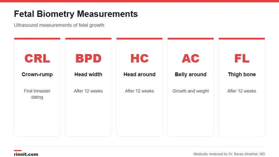
Together these show how the fetus is growing.
Measurement | What it measures | When it's most useful |
CRL (crown-rump length) | Top of the head to the bottom | First trimester, for dating |
BPD (biparietal diameter) | Width of the head | After 12 weeks |
HC (head circumference) | Distance around the head | After 12 weeks |
AC (abdominal circumference) | Distance around the belly | After 12 weeks; reflects growth and weight |
FL (femur length) | Length of the thigh bone | After 12 weeks |
Sources: [1]
Why it's done
Confirm or estimate gestational age and due date.
Check whether the fetus is growing at a healthy rate.
Look for signs of placental problems or maternal conditions.
Monitor pregnancies with complications or twins and other multiples. Sources: [1]
How it's done
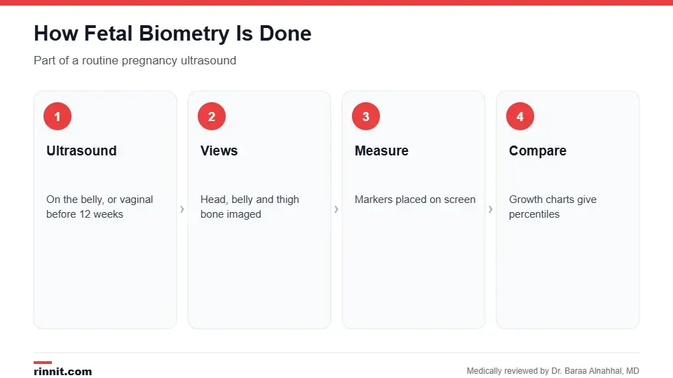
The measurements are part of a routine ultrasound.
You lie on an exam table and gel is spread on your belly. Before 12 weeks, a transvaginal ultrasound may be used.
The sonographer moves the transducer to get clear views of the head, belly and thigh bone.
Measurements are marked on screen and entered into growth charts.
Your provider reviews the results with you. Sources: [1]
Understanding the results
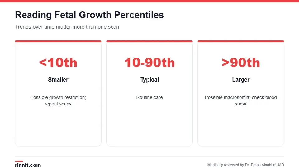
Percentiles compare your baby with others of the same gestational age.
Result | What it may suggest | Usual next step |
10th to 90th percentile | Typical growth | Routine care |
Below 10th percentile | Smaller than average, possible fetal growth restriction or placental problems | Repeat scans, Doppler checks |
Above 90th percentile | Larger than average, possible macrosomia or gestational diabetes | Repeat scans, blood sugar testing |
Your provider looks at growth trends over time rather than a single measurement. Sources: [1, 3]
Accuracy
Biometry is very accurate in early pregnancy, which is why first-trimester CRL is the best way to date a pregnancy. Later in pregnancy, normal differences between babies and measurement error make estimates less precise; estimated fetal weight can be off by a meaningful margin. Sources: [1, 2, 3]
Conclusion
Fetal biometry is a simple, safe part of routine prenatal ultrasound that tracks your baby's growth. It helps your care team date your pregnancy and spot growth concerns early, so they can monitor closely when needed.
Keep learning: read about the pregnancy ultrasound and the 20-week anatomy scan.
Frequently asked questions
What is fetal biometry?
Ultrasound measurements of fetal size used to check growth.
What does BPD mean?
Biparietal diameter, the width of the fetal head.
What does AC measure?
The distance around the fetal belly.
What is CRL?
Crown-rump length, the main measurement in the first trimester.
Is fetal biometry safe?
Yes. It's part of a routine ultrasound with no radiation.
What percentile is normal?
Usually between the 10th and 90th percentiles.
What if my baby measures small?
Your provider will usually repeat scans to check growth over time.
What if my baby measures large?
You may have repeat scans and testing for gestational diabetes.
How accurate is estimated fetal weight?
It's an estimate and can be off, especially late in pregnancy.
When is fetal biometry done?
During routine ultrasounds, often in the first and second trimesters.
Related Reading
Pregnancy Ultrasound: Why It's Done, When You'll Have One, What to Expect and Results the scans where biometry is measured.
20-Week Ultrasound (Anatomy Scan): What It Checks, How to Prepare and What Results Mean the detailed mid-pregnancy scan.
Intrauterine Growth Restriction (IUGR): Causes, Risks, and Management what it means when a fetus is too small.
Fetal Macrosomia: What It Means When Your Baby Is Larger Than Average what it means when a baby is larger than average.
Gestational Diabetes: Symptoms, Causes, Risks & Treatment — Complete Guide a common cause of a larger baby.
References
Cleveland Clinic. Fetal Biometry: Measurements, Normal Values & Accuracy. Cleveland Clinic. Last updated June 20, 2023. https://my.clevelandclinic.org/health/diagnostics/25087-fetal-biometry
Papageorghiou AT, Ohuma EO, Altman DG, et al. International standards for fetal growth based on serial ultrasound measurements: the Fetal Growth Longitudinal Study of the INTERGROWTH-21st Project. Lancet. 2014;384(9946):869-879. PMID 25209488. https://doi.org/10.1016/S0140-6736(14)61490-2
Dudley NJ. A systematic review of the ultrasound estimation of fetal weight. Ultrasound in Obstetrics & Gynecology. 2005;25(1):80-89. PMID 15505877. https://doi.org/10.1002/uog.1751
Medical disclaimer
This article is for general education only and does not replace advice from a qualified healthcare professional. Talk to your doctor about your own diagnosis and treatment, and seek emergency care right away for severe or sudden symptoms.



Comments