Pregnancy Ultrasound: Why It's Done, When You'll Have One, What to Expect and Results

Updated: 2 days ago
Written and medically reviewed by Dr. Baraa Alnahhal, MD · Last reviewed: November 2026 · How we review
Quick Answer: What is a pregnancy ultrasound?
A pregnancy ultrasound, also called a prenatal ultrasound or sonogram, uses sound waves to make pictures of the fetus so a provider can check its growth and development. It can confirm the pregnancy and due date, look for twins and check the placenta and amniotic fluid. The scans have no known harmful side effects.
What the research says
An early routine scan makes twins much less likely to be missed. A Cochrane review of 11 trials with 37,505 women found that routine ultrasound before 24 weeks greatly reduced the chance of failing to detect a multiple pregnancy (risk ratio 0.07) and improved detection of major fetal abnormalities, but it did not change perinatal death rates. (Whitworth et al., Cochrane Database of Systematic Reviews, 2015) [2]
Adding a first-trimester scan may find more structural problems before 24 weeks. A Cochrane review of ultrasound screening in low-risk pregnancies estimated that a first- plus second-trimester scan would correctly identify 83.8% of structural anomalies before 24 weeks, compared with 50.5% for a single second-trimester scan, although no studies compared the two directly. (Buijtendijk et al., Cochrane Database of Systematic Reviews, 2024) [3]
Key takeaways
A pregnancy ultrasound uses sound waves to make images of the fetus and check how it is growing [1].
It can confirm the pregnancy and due date, check the heartbeat, find twins, measure amniotic fluid and locate the placenta [1].
Most people have one or two ultrasounds during pregnancy, and more may be needed in a high-risk pregnancy [1].
An early scan may be done around 7 to 8 weeks, and the anatomy scan is usually done around 18 to 20 weeks [1].
Early scans are often transvaginal, while abdominal scans are generally used after about 12 weeks [1].
Results are either normal or abnormal, and an abnormal or incomplete scan may lead to more scans or tests [1].
What is a pregnancy ultrasound?
A pregnancy ultrasound, also called a sonogram, is a test that checks the growth and development of the fetus [1]. An obstetrician, a nurse midwife or an ultrasound technician called a sonographer may perform it [1].
A small device called a transducer sends sound waves through your belly or vagina, and the echoes are turned into images on a screen [1]. Your provider may order the scan as part of routine care or because they want to look more closely at a possible concern [1].
Pregnancy ultrasounds have no known harmful side effects, but they are still done only when medically needed, with timing based on your health history [1].
Why you might need an ultrasound during pregnancy
Your provider may use an ultrasound to [1]:
Confirm the pregnancy and your due date [1]
Check for early pregnancy complications [1]
Monitor a high-risk pregnancy [1]
Work out the gestational age, or how far along you are [1]
Check fetal growth, movement and heart rate [1]
Look for more than one fetus [1]
Examine your uterus, ovaries and cervix [1]
Measure the amniotic fluid and check where the placenta sits [1]
See the baby's position [1]
Look for problems with the baby's organs, muscles or bones [1]

Ultrasound also guides the needle during amniocentesis or chorionic villus sampling (CVS), and it is part of a biophysical profile, which combines an ultrasound with a nonstress test [1].
When are pregnancy ultrasounds done?
Most people have one or two ultrasounds during pregnancy, although the number and timing depend on your provider and your health [1]. More scans may be suggested if your pregnancy is high risk or a condition is suspected [1].
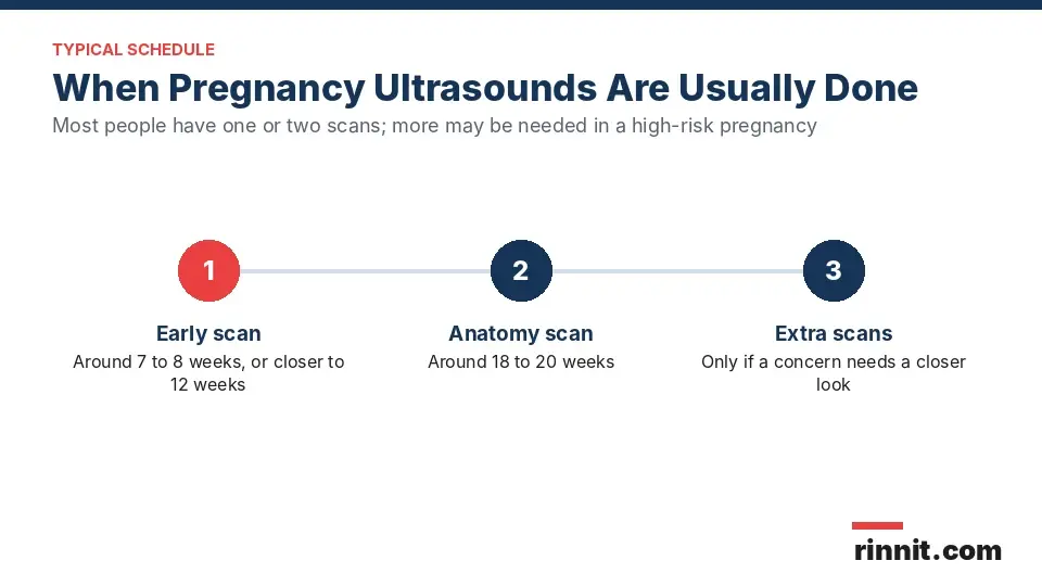
Scan | Usual timing | What it checks |
Early ultrasound | Around 7 to 8 weeks, or closer to 12 weeks with some providers | Confirms the pregnancy by finding a heartbeat, checks for twins, measures the fetus and helps confirm the due date [1] |
Anatomy scan (20-week ultrasound) | Around 18 to 20 weeks | Looks closely at the baby's development; if all is well, it may be the last routine scan [1] |
Extra scans | Any time, if needed | Follow up on a concern, a high-risk pregnancy or an incomplete earlier scan [1] |
Sources: [1]
Types of pregnancy ultrasound
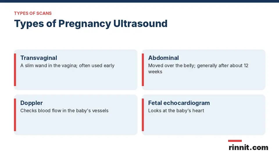
The two main types use the same technology but reach the uterus in different ways [1]:
Your provider may also order special scans:
Doppler ultrasound: Checks blood flow through the baby's blood vessels. Most are done later in pregnancy [1].
Fetal echocardiogram: Looks at the size, shape and function of the baby's heart. It may be used if a heart condition is suspected, if a previous child had a heart condition or if you have certain health conditions [1].
How to prepare for a pregnancy ultrasound
You usually do not need any special preparation, although some providers ask you to come with a full bladder [1]. A support person can come with you, but bringing children is discouraged because the exam needs full focus [1].
For an abdominal scan you usually keep your clothes on and lift your top. For a transvaginal scan in the first trimester, you change into a gown or undress from the waist down [1].
What happens during a pregnancy ultrasound
You lie on a padded table and a gel is spread on your belly [1]. The gel may feel cold, but it does not harm your skin or stain your clothes, and it helps the sound waves travel [1].
The sonographer moves the transducer over your belly, sometimes freezing images or drawing measurement lines on the screen [1]. For a transvaginal scan, the transducer is placed inside the vagina instead, and everything else is the same [1].
After the scan
After an abdominal scan, the gel is wiped off and you may be given pictures to take home [1]. An obstetrician usually reviews the images, often at a follow-up visit the same day [1]. Sonographers usually do not discuss results, but if your obstetrician performs the scan, they may explain what they see as they go [1].
Pregnancy ultrasound results
Results are either normal, meaning no problems were found, or abnormal, meaning something unusual was noticed [1]. An abnormal result usually leads to more ultrasounds or other tests [1].
Sometimes a scan is incomplete because the baby's position or movement makes some areas hard to see, and you may need to come back for a repeat scan [1].
Reasons you may need more ultrasounds
Problems with the ovaries, uterus, cervix or other pelvic organs [1]
A baby who measures small for its age [1]
Placenta problems [1]
Twins, triplets or more [1]
A breech position [1]
Too much or too little amniotic fluid [1]
Conditions such as gestational diabetes or preeclampsia [1]
A congenital disorder in the baby [1]
Conclusion
A pregnancy ultrasound is a safe way to see how your baby is growing and developing. Most people have one scan early in pregnancy and another around 18 to 20 weeks, and some need more if a concern comes up.
Ask your provider which scans you need, when they will be done and who will explain the results to you.
Keep learning: Read our guide to the fetal echocardiogram, our guide to amniocentesis or our due date calculator guide. For more background, see the RadiologyInfo guide to obstetric ultrasound.
FAQ
What is a pregnancy ultrasound?
It is a test that uses sound waves to make pictures of the fetus and check its growth and development.
Is a pregnancy ultrasound safe?
Pregnancy ultrasounds have no known harmful side effects, but they are done only when medically needed.
How many ultrasounds will I have during pregnancy?
Most people have one or two. You may have more if your pregnancy is high risk or a problem is suspected.
When is the first pregnancy ultrasound done?
Some people have an early scan around 7 to 8 weeks. Other providers schedule the first scan closer to 12 weeks.
What is the 20-week ultrasound?
It is the anatomy scan, usually done around 18 to 20 weeks, to look closely at how the baby is developing.
What is the difference between a transvaginal and an abdominal ultrasound?
A transvaginal scan uses a wand inside the vagina and is used early. An abdominal scan moves over the belly and is generally used after about 12 weeks.
Do I need a full bladder for a pregnancy ultrasound?
Usually no special preparation is needed, but some providers ask for a full bladder. Follow your provider's instructions.
Can someone come with me to my ultrasound?
Yes, a support person can come, but bringing children is discouraged because the exam needs full focus.
When will I get my ultrasound results?
An obstetrician usually reviews the images, often at a follow-up visit the same day. Sonographers usually do not discuss results.
Why would I need another ultrasound?
You may need more scans for twins, a breech position, a small baby, placenta or fluid problems, gestational diabetes, preeclampsia or an incomplete scan.
Related Reading
External Cephalic Version (ECV): Turning a Breech Baby explains how ECV turns a breech baby.
Fetal Biometry: Ultrasound Measurements of Baby's Growth Explained explains the ultrasound measurements used to track fetal growth.
Ectopic Pregnancy: Symptoms, Causes, and Treatment explains a pregnancy that grows outside the uterus, an early complication ultrasound helps find.
Intrauterine Growth Restriction (IUGR): Causes, Risks, and Management covers a baby who measures small for its age, a common reason for extra scans.
Breech Baby: What It Means, the Types of Breech Positions, and How Delivery Works explains the breech position that ultrasound can confirm later in pregnancy.
What Is Oligohydramnios? Causes, Signs, and Treatment covers too little amniotic fluid, which ultrasound is used to measure.
Understanding Polyhydramnios: Managing Excess Amniotic Fluid explains too much amniotic fluid, another finding that leads to more scans.
Preeclampsia: Symptoms, Causes & What Helps — A Complete Guide covers a pregnancy condition that often calls for closer ultrasound monitoring.
Biophysical Profile (BPP): Purpose, the Five Parts, Scoring and Results explains the late-pregnancy ultrasound and heart rate test, its five parts and what the score means.
References
Cleveland Clinic. Ultrasound In Pregnancy: What To Expect, Schedule & Results. Cleveland Clinic. Last updated May 11, 2026. https://my.clevelandclinic.org/health/diagnostics/9704-ultrasound-in-pregnancy
Whitworth M, Bricker L, Mullan C. Ultrasound for fetal assessment in early pregnancy. Cochrane Database Syst Rev. 2015;2015(7):CD007058. PMID 26171896. https://doi.org/10.1002/14651858.CD007058.pub3
Buijtendijk MF, Bet BB, Leeflang MM, et al. Diagnostic accuracy of ultrasound screening for fetal structural abnormalities during the first and second trimester of pregnancy in low-risk and unselected populations. Cochrane Database Syst Rev. 2024;5(5):CD014715. PMID 38721874. https://doi.org/10.1002/14651858.CD014715.pub2
Medical disclaimer
This article is for general education only and does not replace advice from a qualified healthcare professional. Talk to your doctor about your own diagnosis and treatment, and seek emergency care right away for severe or sudden symptoms.



Comments