Upper GI Series: What It Is, Why It's Done, How to Prepare and What to Expect

Updated: 2 days ago
Written and medically reviewed by Dr. Baraa Alnahhal, MD · Last reviewed: November 2026 · How we review
Quick Answer: What is an upper GI series?
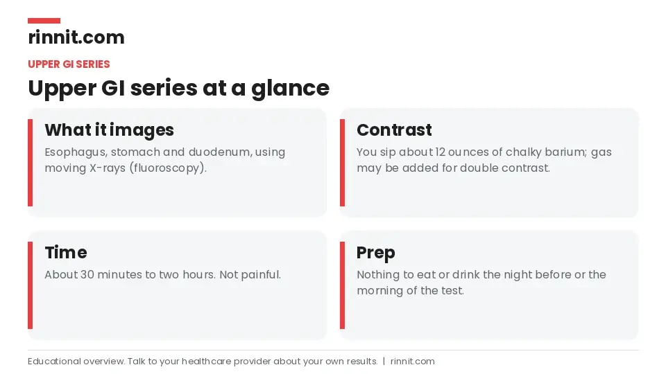
An upper GI series is an imaging test that uses fluoroscopy, a moving form of X-ray, to look inside the esophagus, stomach and the first part of the small intestine. You drink a chalky liquid called barium that coats these organs so they stand out. It's painless and helps find tissue changes, structural problems and swallowing or emptying problems. [1]
What the research says
Barium studies spot most stomach cancers, but cannot confirm them. In 60 people with proven stomach cancer, the upper GI series detected 95% of tumors (57 of 60), close to endoscopy at 98%. A tissue sample is still needed to confirm cancer. (Chen et al., World Journal of Gastroenterology, 2003) [2]
Mild reflux damage is easy to miss on any imaging. In 37 people with reflux symptoms, double-contrast barium studies picked up reflux esophagitis in 35% of biopsy-proven cases, similar to endoscopy at 39%, so a normal result does not fully rule out mild inflammation. (Dibble et al., Abdominal Imaging, 2004) [3]
Key takeaways
An upper GI series uses fluoroscopy and a barium drink to show your esophagus, stomach and duodenum in motion. [1]
It helps find inflammation, ulcers, hiatal hernia, narrowing, swallowing problems and slow stomach emptying. [1]
You need an empty stomach, so you usually stop eating and drinking the night before. [1]
The test takes 30 minutes to two hours and is not painful. [1]
Constipation from leftover barium is the most common side effect, so drink plenty of water afterward. [1]
It can show a tumor but cannot tell if it is cancer; that needs a biopsy during an upper endoscopy. [1]
What is an upper GI series?
Your gastrointestinal (GI) tract is a long, hollow tube that runs from your mouth to your anus. "Upper GI" means the top part of this tube: your esophagus (swallowing pipe), your stomach and your duodenum. [1]
The "series" is a set of fluoroscopic X-rays taken during the test. A regular X-ray is a still picture. Fluoroscopy is more like a short video, so your provider can watch your organs working and see more detail on their inner surface. [1]
How it fits with other barium tests
Providers use several barium X-ray exams, each focused on a different part of the GI tract. They may order one or more of them depending on where your symptoms come from. [1]
Test | What it examines |
Esophagram (barium swallow) | The esophagus only |
Upper GI series | Esophagus, stomach and duodenum |
Small bowel series (small bowel follow-through) | The small intestine |
Lower GI series (barium enema) | The lower small intestine (ileum) and the large intestine |
Sources: [1]
The esophagus, stomach and duodenum are grouped together because a swallowed liquid passes through all three fairly quickly, this is about as far as an instrument can reach from the top, and diseases in one part often affect the others. [1]
Why is an upper GI series done?
An upper GI series is often the first step in finding the cause of unexplained symptoms in the upper GI tract. Your provider may order it if you have: [1]
Swallowing problems
Heartburn
Abdominal pain
Nausea and vomiting
Indigestion
What can an upper GI series detect?
Type of problem | Examples |
Tissue changes | Gastritis, esophagitis, erosion from acid or bile reflux, peptic ulcer disease, Barrett's esophagus, gastric intestinal metaplasia |
Structural problems | Esophageal varices, esophageal strictures (narrowing), hiatal hernia, polyps, tumors |
Functional problems | Dysphagia, gastroparesis, achalasia, laryngopharyngeal reflux, pyloric valve problems, blockages |
Sources: [1]

Can it detect acid reflux?
Chronic acid reflux (GERD) is often diagnosed from symptoms alone. An upper GI series can still show signs that point to GERD, such as severe acid erosion, ulcers or strictures caused by reflux, or a sphincter muscle that does not close all the way. [1]
Can it detect cancer?
It can show tumors and tissue changes if they are large enough, but it cannot tell whether they are cancer. To diagnose cancer, your provider takes a tissue sample (biopsy) during an upper endoscopy. [1]
Upper GI series vs. upper endoscopy
Both tests look at the same organs, but they work very differently. [1]
Feature | Upper GI series | Upper endoscopy (EGD) |
How it works | Fluoroscopic X-rays with a barium drink | A thin camera passed down your throat |
Invasive? | No | Yes, often with sedation |
Can take a biopsy? | No | Yes |
Typical role | Often the first test | Often a follow-up to imaging |
Sources: [1]
How does the test work?
X-rays and fluoroscopy. X-rays pass through your body, and dense parts look brighter while soft tissues look gray. Fluoroscopy sends a continuous beam for seconds or minutes to make a short video, which means slightly more radiation than a single X-ray, but a level considered safe in most cases. [1]
Barium contrast. Barium is a thick, white, chalky liquid that coats the inside of your organs so they show up clearly. Your body does not absorb it, and it passes out in your stool. [1]
Double contrast. Sometimes gas is added as a second contrast agent. You swallow a tablet or a fizzy drink, and the gas gently stretches the organ walls. Gas looks black on X-ray, which sharpens the contrast with the white barium. [1]
How do you prepare for an upper GI series?
Before scheduling: Tell your provider about any recent X-rays or other radiation exposure, since they may want to space out your tests. [1]
Pregnancy: Tell your provider if you are or might be pregnant. Radiation exposure to a fetus is linked to birth defects, so they will plan another approach. [1]
Fasting: Do not eat or drink anything the night before or the morning of the test, so your stomach is empty. [1]
Medicines: You may be asked to stop some medicines for a short time, such as antacids. [1]
What happens during the test?
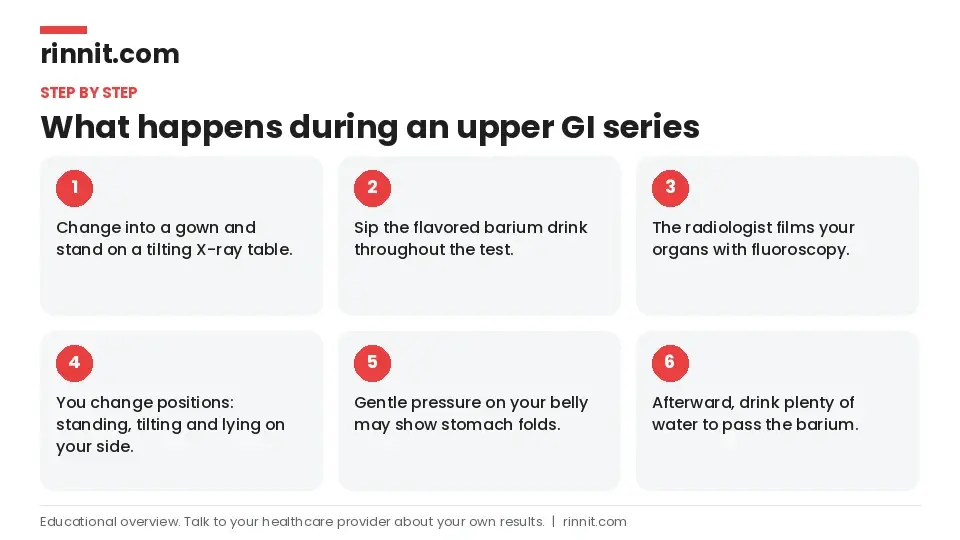
You change into a hospital gown and go to a room with a tilting X-ray table. You start standing, and a technologist secures you with straps. [1]
You sip a sweetened, flavored barium drink throughout the test, about 12 ounces in total. Some parts may use a thicker or thinner version, and you may also swallow gas-producing crystals or a fizzy drink. [1]
The radiologist takes pictures from different angles. You may stand at a 45-degree angle, tilt back, lie on one side, or roll slightly against a sponge so the barium reaches every area. [1]
The radiologist may gently press on your belly to show the folds of your stomach. [1]
The test takes 30 minutes to two hours. It can take longer if the barium moves slowly through your duodenum. [1]
Does it hurt?
The test itself is not painful, and you can ask for a sedative if you want one. Afterward you may feel a little bloated or crampy, and you may have some gas pain if you had a double-contrast study. These feelings usually settle within a day or so. [1]
What are the risks and side effects?
Risk | What to know |
Constipation | The most common side effect. Drink plenty of water. Your stool may be white for a day or so. Call your provider if you cannot poop; they may prescribe a laxative. |
Kidney complications | People with advanced kidney disease face a risk of internal bleeding in the upper GI tract, and the contrast may affect kidney function. |
Radiation | Effects add up over a lifetime. One fluoroscopic exam is safe for most people who are not pregnant. |
Allergy | A small risk of an allergic reaction to substances in the contrast drink. |
Sources: [1]
Who should not have an upper GI series?
This test may not be a good option if you: [1]
Are pregnant
Have kidney disease
Already have constipation
Have recently been exposed to radiation from other sources
Have a known or suspected hole (perforation) in your upper GI tract
Have swallowing problems that cause food or liquid to go into your lungs (aspiration)
What do the results mean?
A radiologist studies your images and sends a report to your healthcare provider, who goes over the results with you. Some conditions can be diagnosed from the X-rays alone. Others need follow-up tests, such as an upper endoscopy with a biopsy. [1]
Conclusion
An upper GI series is a painless, noninvasive way to look inside your esophagus, stomach and duodenum. Because fluoroscopy shows your organs in motion, your provider can see both how they look and how well they work. [1]
If the images do not give a final answer, they usually point to the right next step. Ask your provider what they are looking for and how your results will guide your care.
Keep learning: Read our guide to the esophagram (barium swallow) or our guide to acid reflux and GERD. For more trusted, doctor-reviewed health guides, visit Rinnit.com.
FAQ
What is an upper GI series?
It is an X-ray test that uses fluoroscopy and a barium drink to look at your esophagus, stomach and duodenum. [1]
Why would a doctor order an upper GI series?
To look for the cause of swallowing problems, heartburn, belly pain, nausea, vomiting or indigestion. [1]
What can an upper GI series show?
It can show inflammation, ulcers, hiatal hernia, narrowing, polyps, tumors, swallowing problems and slow stomach emptying. [1]
How do I prepare for an upper GI series?
Do not eat or drink the night before or the morning of the test, and ask if you should pause medicines such as antacids. [1]
How long does an upper GI series take?
It usually takes 30 minutes to two hours. [1]
Is an upper GI series painful?
No. You may feel bloated or crampy afterward, which usually passes in a day or so. [1]
What are the side effects of an upper GI series?
Constipation is the most common. Drinking lots of water helps the barium pass. [1]
Can an upper GI series detect cancer?
It can show a tumor, but a biopsy during an upper endoscopy is needed to confirm cancer. [1]
Who should not have an upper GI series?
People who are pregnant or have kidney disease, constipation, a GI perforation or aspiration may need a different test. [1]
When should I call my doctor after an upper GI series?
Call if you cannot have a bowel movement after the test, since you may need a laxative. [1]
Related Reading
Gastric Emptying Study in Children (Solid-Meal GES): What to Expect explains the stomach emptying test for children.
Contrast Enema for Children: What to Expect for Your Child explains the X-ray test that views a child's colon.
Gastrointestinal (GI) Tests: Types, Preparation and What to Expect explains the main types of tests used to check the digestive tract.
Enteroscopy: Seeing and Treating the Small Intestine explains the scope exam that reaches deep into the small intestine.
Acid Reflux & GERD: Symptoms, Causes, Diagnosis, and Treatment explains chronic acid reflux, which can leave erosion, ulcers or strictures that an upper GI series may show.
Peptic Ulcer Disease: Causes, Symptoms, Diagnosis, and Treatment covers stomach and duodenal ulcers, one of the tissue changes this test can reveal.
Hiatal Hernia: Symptoms, Causes, and Treatment Options describes the structural problem in which part of the stomach pushes up through the diaphragm.
Achalasia: Symptoms, Causes, and Treatment explains the esophageal sphincter problem that fluoroscopy can show in action.
Dysphagia (Trouble Swallowing): Symptoms, Causes, Diagnosis & Treatment — Complete Guide covers trouble swallowing, a common reason to order this test.
Esophageal Strictures: Symptoms, Causes, Diagnosis, Treatment and When to Seek Emergency Care — What You Need to Know explains narrowing of the esophagus, which barium can outline.
Gastroparesis: Symptoms, Causes, Diet & Treatment — Complete Guide describes slow stomach emptying, a motility problem the test can help detect.
Esophagitis: Symptoms, Causes, and Treatment covers inflammation of the esophagus, one of the tissue changes seen on an upper GI series.
Gastric Emptying Study: How to Prepare, What Happens and Normal Results explains the scan that measures how fast food leaves the stomach.
References
Cleveland Clinic. Upper GI Series: Definition, Procedure & Tests. Cleveland Clinic. Last updated February 14, 2022. https://my.clevelandclinic.org/health/diagnostics/22451-upper-gi-series
Chen F, Ni YC, Zheng KE, et al. Spiral CT in gastric carcinoma: comparison with barium study, fiberoptic gastroscopy and histopathology. World J Gastroenterol. 2003;9(7):1404-1408. PMID 12854130. https://doi.org/10.3748/wjg.v9.i7.1404
Dibble C, Levine MS, Rubesin SE, et al. Detection of reflux esophagitis on double-contrast esophagrams and endoscopy using the histologic findings as the gold standard. Abdom Imaging. 2004;29(4):421-425. PMID 14716454. https://doi.org/10.1007/s00261-003-0128-5
Medical disclaimer
This article is for general education only and does not replace advice from a qualified healthcare professional. Talk to your doctor about your own diagnosis and treatment, and seek emergency care right away for severe or sudden symptoms.



Comments