Transesophageal Echocardiogram (TEE): Why It's Done, What to Expect and Risks

Updated: 2 days ago
Written and medically reviewed by Dr. Baraa Alnahhal, MD · Last reviewed: November 2026 · How we review
Quick Answer: What is a transesophageal echocardiogram?
A transesophageal echocardiogram (TEE) is an ultrasound test that makes detailed pictures of the heart and upper aorta from inside the body. While you're sedated, a provider guides a thin, flexible tube down the throat into the esophagus, right behind the heart. It can find valve disease, blood clots, infections and aortic aneurysms. You'll need a ride home. [1]
What the research says
TEE sees heart infections that a regular echo can miss. A meta-analysis of 16 studies (2,807 patients) used TEE as the reference standard and found that a modern transthoracic echo detected only 61% of the growths (vegetations) seen on TEE, though it was 94% specific. The authors conclude a follow-up TEE is almost always needed unless a regular echo is clearly negative. (Bai et al., Journal of the American Society of Echocardiography, 2017) [2]
A clearly negative regular echo can help rule out infection in low-risk patients. Another meta-analysis of 11 studies (2,209 patients) with suspected native valve endocarditis found transthoracic echo had 71% sensitivity and 80% specificity compared with TEE. (Bonzi et al., Internal and Emergency Medicine, 2018) [3]
Key takeaways
A TEE takes ultrasound pictures of your heart from inside your esophagus.
It gives a much clearer view of the back of the heart than a standard echo.
You'll be sedated, awake but drowsy, and your throat is numbed.
Stop eating and drinking usually at least 6 hours before the test.
You can't drive for 24 hours afterward, and a sore throat is common.
What is a TEE?
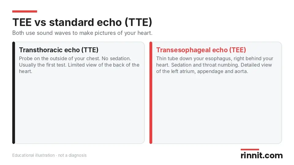
A TEE uses sound waves to make detailed pictures of your heart and upper aorta. Unlike a transthoracic echo (TTE), which is done from outside your chest, a TEE takes pictures from inside your body. [1]
Your provider guides a long, thin tube called an endoscope down your esophagus. A transducer on its tip sends sound waves that bounce off your heart, and a computer turns the echoes into pictures of its structure and function. [1]
Why the esophagus?
Your esophagus runs right behind your heart, so it's the perfect spot to send and receive sound waves. A TEE is especially valuable for seeing structures at the back of your heart: [1]
Left atrium: the top left chamber of your heart
Left atrial appendage (LAA): a small, ear-shaped pouch where blood clots can form
Interatrial septum: the wall between your left and right atria
Thoracic aorta: the part of your aorta that runs through your chest
Your provider may add Doppler and color Doppler, which show the speed and direction of blood flow through your heart. [1]
Why would you need a TEE?
You may need a TEE if: [1]
Other tests didn't show enough detail. A TTE may not have been enough to diagnose or manage a heart problem.
You're in critical condition. It helps if you have very low blood pressure, low oxygen or shock, or sudden aortic problems such as a tear, chest trauma or prosthetic valve issues.
You're having a procedure for an arrhythmia. Before catheter ablation or electrical cardioversion, a TEE checks for heart clots that could break free.
You're having heart surgery. It can check your anatomy before surgery, confirm success afterward and guide some procedures in real time.
Providers usually do a TTE first because it's less invasive. They may go straight to TEE if you have chest anatomy that makes a TTE hard, lung disorders or a life-threatening emergency. [1]
Feature | Transthoracic echo (TTE) | Transesophageal echo (TEE) |
Where the probe goes | On the outside of your chest | Down your throat into your esophagus |
Sedation | Not needed | Yes, usually moderate sedation |
View of back of heart | Limited | Detailed |
Typical use | First test | When more detail is needed, or before and during some procedures |
Sources: [1]
How do you prepare for a TEE?
Your provider will tell you: [1]
When to stop eating and drinking (usually at least 6 hours before)
When to take your usual medicines
When to avoid alcohol before the test
Anything else to do or avoid
Tell your provider about these conditions
Some conditions make a TEE riskier, though they don't always rule it out. Tell your provider if you have: [1]
Esophageal or stomach cancer
Gastritis or GERD (chronic acid reflux)
A hiatal hernia
Problems swallowing
Sleep apnea
A history of IV drug use
Also mention any medicines you take for sleep, anxiety or pain. [1]
What happens during a TEE?
You put on a hospital gown.
Small stickers (electrodes) go on your chest to track your heart's electrical activity.
A blood pressure cuff goes on your arm and a pulse oximeter on your finger.
Your throat is sprayed with medicine to prevent pain and gagging.
You get a sedative through an IV to help you relax, and possibly oxygen through your nose.
You lie on your left side.
Your provider gently guides the lubricated tube through your mouth and down your throat, and may ask you to swallow.
They take pictures; you may feel some pressure in your chest but shouldn't feel pain.
They remove the tube and keep watching your blood pressure, pulse and oxygen.
Sources: [1]
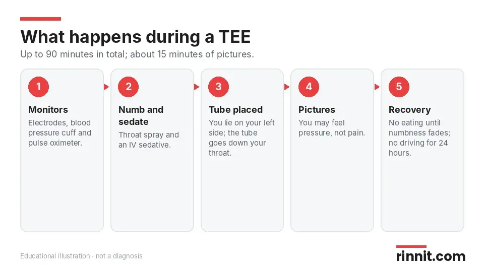
The whole visit may take up to 90 minutes, but the picture-taking usually takes only about 15 minutes. [1]
Are you awake during a TEE?
You're awake but drowsy. Most people don't need general anesthesia. If you do have it, you'll be fully asleep and won't remember the test, but recovery may take longer. [1]
What are the risks of a TEE?
A TEE is an invasive test but not surgery, and the risk of serious problems is low. Rarely, the tube can injure or tear the throat or esophagus, so call your provider for chest pain, trouble swallowing or vomiting blood afterward. Possible complications include: [1]
Allergic reactions to medicines
Aspiration pneumonia
Blood pressure or heart rhythm problems
Minor bleeding in your esophagus
What to expect after a TEE
Someone must drive you home, because you can't drive for 24 hours after sedation. You can expect to: [1]
Have a numb throat for about an hour. Don't eat or drink until it wears off, or you might choke.
Have a sore throat. It may last a few hours to a few days. Call your provider if it doesn't go away or gets worse.
Your provider will tell you when your results will be ready. [1]
What can a TEE show?
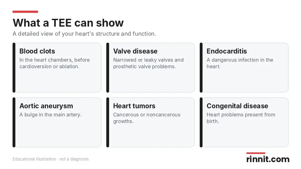
A TEE gives a detailed view of your heart's structure and function. It can help diagnose: [1]
Condition | What the TEE can show |
Aortic aneurysm | A bulge in the artery carrying blood away from your heart |
Blood clots | Clumps of blood inside your heart chambers |
Congenital heart disease | Heart problems you're born with |
Heart tumors | Growths in the heart, cancerous or not |
Heart valve disease | Narrowed or leaky valves, or growths or infection on prosthetic valves |
Infective endocarditis | A dangerous infection in your heart |
Pericarditis | Inflammation of the sac around your heart |
Sources: [1]
Questions to ask your provider
Why do I need a TEE instead of a regular echo?
When should I stop eating, drinking and taking my medicines?
Will I have moderate sedation or general anesthesia?
When will I get my results?
What symptoms after the test should make me call you?
Conclusion
A transesophageal echocardiogram gives your care team a close-up view of your heart that a standard echo can't always provide. It is especially useful for checking for clots, infections and valve problems, and for guiding certain procedures.
The idea of a tube down your throat can feel unsettling, but you'll be numbed and sedated, and the picture-taking part is short. Plan for a ride home and expect a sore throat for a day or two.
Keep learning: Read our guides to the echocardiogram, the fetal echocardiogram, Doppler ultrasound and endoscopy. For more, see American Heart Association: Transesophageal Echocardiography.
FAQ
What is a transesophageal echocardiogram?
It is an ultrasound test that takes detailed pictures of your heart from inside your esophagus. [1]
Why would a doctor order a TEE?
When a regular echo isn't detailed enough, in critical illness, before arrhythmia procedures or during heart surgery. [1]
Are you awake during a TEE?
You're awake but drowsy from sedation; most people don't need general anesthesia. [1]
Does a TEE hurt?
Your throat is numbed and you're sedated, so you shouldn't feel pain, though you may feel pressure. [1]
How long does a TEE take?
Up to 90 minutes in total, with about 15 minutes of picture-taking. [1]
Do I need to fast before a TEE?
Yes, usually for at least 6 hours, but follow your provider's instructions. [1]
Can I drive after a TEE?
No. You can't drive for 24 hours because of the sedation. [1]
Is a TEE major surgery?
No. It is an invasive diagnostic test, not surgery, and serious problems are uncommon. [1]
What are the risks of a TEE?
Allergic reactions, aspiration pneumonia, blood pressure or heart rhythm problems and minor bleeding in the esophagus. [1]
When can I eat after a TEE?
Wait until the numbness in your throat wears off, usually about an hour. [1]
Related Reading
Learn more in our guide to Endocarditis: Symptoms, Causes, Types, Diagnosis, Treatment and When to Seek Emergency Care — What You Need to Know.
Learn more in our guide to Heart Valve Disease: Symptoms, Causes & Treatment.
Learn more in our guide to Aortic Aneurysm: Types, Causes, Dissection Risk & Complete Guide.
Learn more in our guide to What Is a Cardiac Tumor? A Complete Guide to Heart Tumors, Types, and Treatments.
Learn more in our guide to Congenital Heart Disease (CHD): Types, Symptoms, Treatment & Survival — Complete Guide.
Learn more in our guide to Pericarditis: Symptoms, Causes & Treatment — Why Sharp Chest Pain Needs a Prompt Check.
Learn more in our guide to Heart Arrhythmia: Symptoms, Types, Causes, Diagnosis & Treatment — Complete Guide.
Bubble Study (Bubble Echo): Finding PFO, ASD and Lung Shunts explains the saline bubble ultrasound test that finds holes in the heart.
References
Cleveland Clinic. Transesophageal Echocardiogram (TEE). Cleveland Clinic. Last updated July 15, 2025. https://my.clevelandclinic.org/health/diagnostics/4992-echocardiogram-transesophageal-tee
Bai AD, Steinberg M, Showler A, et al. Diagnostic Accuracy of Transthoracic Echocardiography for Infective Endocarditis Findings Using Transesophageal Echocardiography as the Reference Standard: A Meta-Analysis. J Am Soc Echocardiogr. 2017;30(7):639-646.e8. PMID 28483353. https://doi.org/10.1016/j.echo.2017.03.007
Bonzi M, Cernuschi G, Solbiati M, et al. Diagnostic accuracy of transthoracic echocardiography to identify native valve infective endocarditis: a systematic review and meta-analysis. Intern Emerg Med. 2018;13(6):937-946. PMID 29546685. https://doi.org/10.1007/s11739-018-1831-0
Medical disclaimer
This article is for general education only and does not replace advice from a qualified healthcare professional. Talk to your doctor about your own diagnosis and treatment, and seek emergency care right away for severe or sudden symptoms.



Comments