Lymphoscintigraphy (Lymph Node Mapping): Uses, Procedure and Results

Updated: 2 days ago
Written and medically reviewed by Dr. Baraa Alnahhal, MD · Last reviewed: November 2026 · How we review
Quick Answer
Lymphoscintigraphy (lymph node mapping) is a nuclear medicine imaging test that shows how lymph fluid flows through the lymphatic system. A radioactive tracer is injected under the skin, and a gamma camera follows it to nearby lymph nodes. It's often used before surgery for breast cancer and melanoma to find the sentinel lymph node and to diagnose lymphedema.
What the research says
Different injection sites work for sentinel node mapping: a meta-analysis in breast cancer found both superficial (near the skin) and deep (near the tumor) injections of tracer and blue dye were effective for finding axillary sentinel nodes on lymphoscintigraphy and at surgery, while deep injections found nodes outside the armpit three times more often (Ahmed M et al., British Journal of Surgery, 2015) [2]
Mapping still works after previous surgery: in a meta-analysis of people with recurrent breast cancer, lymphatic mapping found a sentinel node in 59.6% and unusual drainage routes in 25.7%, helping more than half of those with prior sentinel node biopsy avoid a full lymph node removal (Ahmed M et al., British Journal of Surgery, 2016) [3]
Key takeaways
Maps lymph flow using a radiotracer injected under the skin and a gamma camera.
Used to find sentinel nodes in breast cancer and melanoma, diagnose lymphedema and check for blocked lymph vessels.
Remove jewelry and tell your team if you're pregnant or breastfeeding.
The injection may sting briefly; the scan itself doesn't hurt and takes 30 minutes to a few hours.
The tracer leaves your body in urine and stool over a couple of days.
What is lymphoscintigraphy?
Your lymphatic system is a network of vessels and nodes that drains fluid from tissues and helps fight infection. Cancer cells can travel through it to nearby lymph nodes. Lymphoscintigraphy uses a radiotracer that's absorbed into lymph vessels, so the gamma camera can show where lymph flows, where it collects and whether flow is blocked. Sources: [1]

Lymphoscintigraphy follows a tracer through the lymph vessels.
Why is it done?
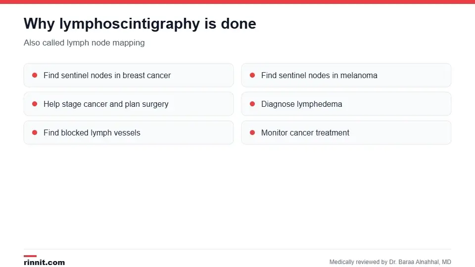
Uses of lymph node mapping.
Sentinel node mapping: to find the first lymph node(s) draining a breast cancer or melanoma so the surgeon can remove and test them.
Cancer staging and surgical planning: to show whether cancer may have spread and guide a biopsy.
Lymphedema: to diagnose swelling caused by poor lymph drainage, including genital lymphedema, and to find blocked lymph vessels.
Treatment monitoring: to check whether cancer treatment is working. Sources: [1]
How do I prepare?
Remove jewelry and other metal.
Tell your provider if you are pregnant or breastfeeding.
Bring a list of your medicines, vitamins and supplements.
You may change into a gown. Sources: [1]
If the scan is done to find sentinel nodes, it's usually on the same day as your surgery. You generally don't need to stay in the hospital for the scan itself. Sources: [1]
What happens during the test?
A small amount of radiotracer is injected just under the skin near the tumor, scar or swollen area; you may feel a brief sting.
The tracer moves through your lymph vessels toward nearby nodes.
You lie on a table while a gamma camera takes a series of images; it may stay still or rotate around you.
You may need to change positions and hold still for short periods. Sources: [1]
The test can take about 30 minutes or a few hours, depending on the reason. Sources: [1]
Sentinel node biopsy

How lymph node mapping leads to a sentinel node biopsy.
In breast cancer and melanoma, the images show the surgeon which node(s) to remove. During surgery, a handheld probe detects the tracer, sometimes with a blue dye, and the sentinel node is sent to the lab. If it's free of cancer, you may be able to avoid removing more nodes, such as an axillary lymph node dissection, which can cause lymphedema. Sources: [1, 2, 3]
After the test
You can usually go home and return to normal activities right away. The tracer leaves your body through urine and stool over the next couple of days; drinking plenty of water helps. Sources: [1]
Risks
The radiation dose is low. Side effects are rare, though some people have an allergic reaction to the tracer. The test may not be suitable during pregnancy or breastfeeding, so tell your team beforehand. Sources: [1]
Understanding your results
A radiologist reviews the images and sends a report to your provider, usually within a week. If the scan suggests cancer has spread, the next step is typically a sentinel node biopsy. If it shows poor lymph drainage, your provider may recommend lymphedema treatment such as compression and lymphatic drainage massage. Sources: [1]
Conclusion
Lymphoscintigraphy shows exactly where lymph flows, which helps surgeons remove only the lymph nodes that matter and helps diagnose lymphedema. It's safe, usually same-day and involves only a quick injection. Ask whether your scan is for mapping before surgery or for checking swelling, so you know what to expect.
Keep learning: read about the lymphatic system and lymph node removal.
Frequently asked questions
What is lymphoscintigraphy used for?
To find sentinel lymph nodes in cancer, diagnose lymphedema and check lymph flow.
Is lymphoscintigraphy painful?
The injection may sting briefly; the scan itself doesn't hurt.
How long does it take?
About 30 minutes to a few hours.
Is it done on the day of surgery?
Often, when it's used to find sentinel nodes.
Is the radiation dangerous?
The dose is low, but tell your team if you're pregnant or breastfeeding.
What is a sentinel lymph node?
The first lymph node that cancer cells would spread to from a tumor.
Can I go home after the test?
Yes, usually right away.
How long does the tracer stay in my body?
It leaves through urine and stool over a couple of days.
When will I get results?
Usually within a week.
What happens if results are abnormal?
Usually a sentinel node biopsy, or lymphedema treatment if drainage is poor.
Related Reading
Understanding the Lymphatic System: Your Body's Silent Guardian the system this scan maps.
Axillary Lymph Node Dissection: Understanding the Procedure, Risks, and Recovery surgery a negative sentinel node can help avoid.
Lymphadenectomy: What Lymph Node Removal Involves, Recovery and Risks removal of lymph nodes in cancer surgery.
Genital Lymphedema: Symptoms, Causes, Diagnosis, Treatment, and Outlook — What You Need to Know swelling diagnosed with lymph imaging.
Melanoma: A Comprehensive Guide to Early Detection and Treatment a cancer where sentinel node mapping is used.
Sentinel Node Biopsy: How It Works, Recovery, Risks and Results explains the biopsy that checks whether cancer has reached the first lymph nodes.
Lymphangiography: Imaging the Lymphatic System for Leaks and Lymphoma explains the dye imaging test that maps lymph vessels and nodes.
References
Cleveland Clinic. Lymphoscintigraphy: Procedure Details & Results. Cleveland Clinic. Last updated March 24, 2026. https://my.clevelandclinic.org/health/diagnostics/lymphoscintigraphy
Ahmed M, Purushotham AD, Horgan K, et al. Meta-analysis of superficial versus deep injection of radioactive tracer and blue dye for lymphatic mapping and detection of sentinel lymph nodes in breast cancer. Br J Surg. 2015;102(3):169-181. PMID 25511661. https://doi.org/10.1002/bjs.9673
Ahmed M, Baker R, Rubio IT. Meta-analysis of aberrant lymphatic drainage in recurrent breast cancer. Br J Surg. 2016;103(12):1579-1588. PMID 27598038. https://doi.org/10.1002/bjs.10289
Medical disclaimer
This article is for general education only and does not replace advice from a qualified healthcare professional. Talk to your doctor about your own diagnosis and treatment, and seek emergency care right away for severe or sudden symptoms.



Comments