Lymphangiography: Imaging the Lymphatic System for Leaks and Lymphoma

Updated: 3 days ago
Written and medically reviewed by Dr. Baraa Alnahhal, MD · Last reviewed: November 2026 · How we review
Quick Answer
Lymphangiography (lymphography) is an imaging test that uses injected dye with X-ray or MRI to show the lymphatic vessels and lymph nodes, which regular imaging can't see well. It's used to find lymphatic leaks, evaluate lymphedema, check lymphoma and map lymph vessels before surgery. Your skin, urine and stool may look discolored for a while afterward.
What the research says
Lymphangiography often guides treatment of chyle leaks: in 109 people with high-output chyle leaks after thoracic duct injury, lymphangiography was successful in 108, and when the duct could be reached and blocked, the leak resolved in 90% of those treated (Itkin M et al., J Thorac Cardiovasc Surg, 2010) [2]
Lymph node injection approaches work well: in nine people with a chyle leak after thoracic outlet surgery, ultrasound-guided intranodal lymphangiography with thoracic duct embolization achieved clinical success in 8 of 9, with no recurrence during follow-up (Kim SK et al., J Vasc Interv Radiol, 2020) [3]
Key takeaways
Uses dye with X-ray or MRI to map lymph vessels and nodes.
Finds lymphatic leaks, lymphedema and lymphoma spread.
Can guide treatment, sometimes during the same procedure.
Dye flows in for about an hour through a small tube.
Skin, urine and stool may look discolored afterward.
What is the lymphatic system?
The lymphatic system is a network of vessels and lymph nodes that carries fluid and helps fight infection. Its vessels are tiny and clear, so they don't show up well on routine scans. Lymphangiography uses dye to make them visible. When MRI is used, it's called MR lymphangiography. Sources: [1]
Why is it done?
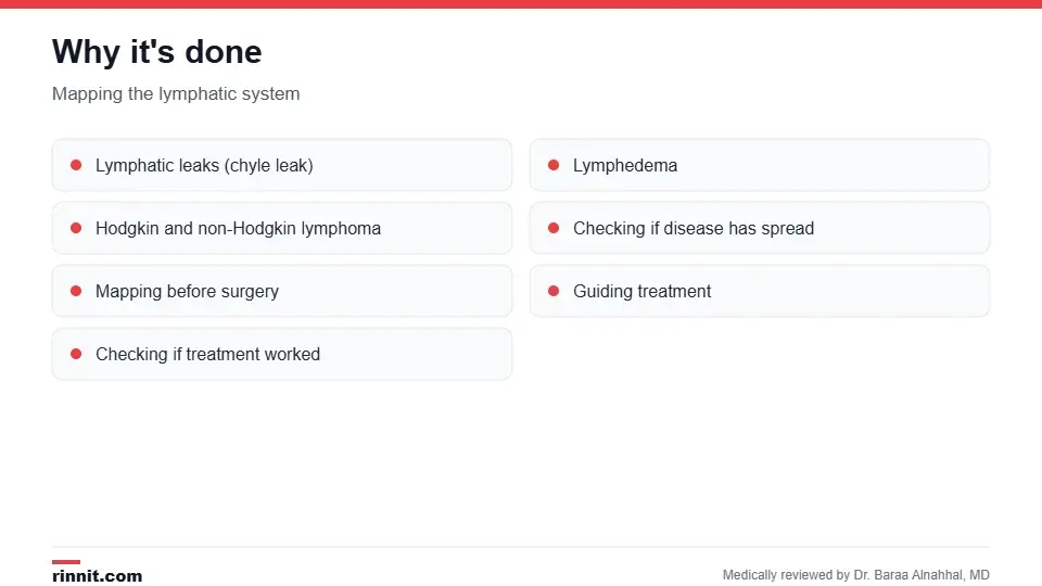
Lymphangiography maps the lymphatic system.
To find lymphatic leaks or injuries, such as a chyle leak or chylothorax.
To evaluate lymphedema.
To help diagnose and stage Hodgkin lymphoma and non-Hodgkin lymphoma.
To see whether disease has spread.
To map lymph vessels before surgery and guide treatment, and to check whether treatment worked. Sources: [1, 2]
A related nuclear test, lymphoscintigraphy, also maps lymph flow. Sources: [1]
How do I prepare?
Don't eat or drink for several hours beforehand, as instructed.
Empty your bladder just before the test.
Tell your provider if you could be pregnant, have bleeding problems, have allergies, including to iodine, or have heart conditions such as a patent foramen ovale.
You may get medicine to help you relax. Sources: [1]
What happens during the test?
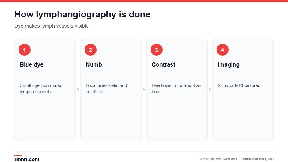
Contrast flows in for about an hour.
You sit or lie down, and the skin is cleaned, usually near the groin, or between the fingers or toes.
A small amount of blue dye is injected to outline the lymph channels.
One area is numbed, a small cut is made and a thin, flexible tube is inserted, sometimes guided by ultrasound.
Contrast dye is injected slowly for about an hour; you may feel pressure.
Fluoroscopy or MRI images confirm the dye is moving.
The tube is removed, the cut is stitched, and images of the legs, pelvis, belly and chest are taken, sometimes again the next day. Sources: [1]
Risks
Lymphangiography is generally safe. It uses a small amount of radiation when X-rays are used. Possible complications include an allergic reaction, bleeding, infection and inflammation of the lymph vessels. Sources: [1]
Recovery
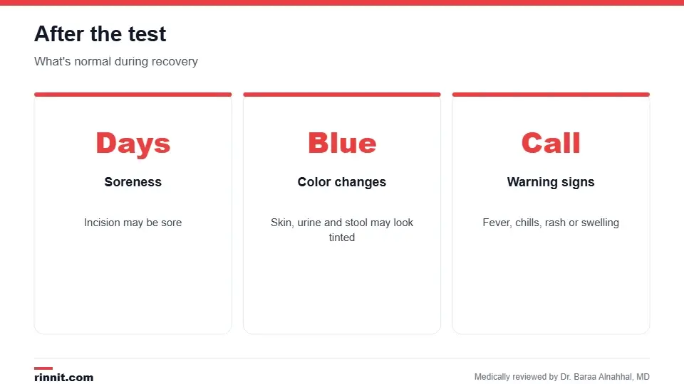
Dye can stay in lymph nodes for a long time.
The cut may be sore for a few days. Your skin, urine and stool may look discolored until most of the dye leaves your body, and some dye can stay in lymph nodes for up to two years. Call your provider for fever, chills, pain or a red, swollen rash. Sources: [1]
Understanding your results
Results can often be read the same day, and treatment is sometimes done during the same procedure. A blockage where dye can't pass may point to a tumor, injury or infection. Enlarged, foamy-looking lymph nodes may suggest lymphatic cancer. An interventional radiologist and oncologist may review results together. Sources: [1, 2, 3]
Conclusion
Lymphangiography makes the hidden lymphatic system visible. It's especially valuable for finding and treating lymph leaks and for mapping the lymph system before surgery or in lymphoma.
Keep learning: read about the lymphatic system and chylothorax.
Frequently asked questions
What is lymphangiography?
An imaging test that uses dye to show lymph vessels and nodes.
Why is it done?
To find lymph leaks, lymphedema, lymphoma and disease spread.
Does it hurt?
The area is numbed; you may feel a sting and pressure.
How long does it take?
Dye is injected for about an hour, plus imaging time.
Will my skin change color?
Skin, urine and stool may look discolored for a while.
Is there radiation?
A small amount when X-rays are used; MRI uses none.
Can treatment be done at the same time?
Sometimes, such as blocking a leaking lymph duct.
Do I need to fast?
Usually, for several hours beforehand.
When will I get results?
Often the same day.
What symptoms should I report afterward?
Fever, chills, pain or a red, swollen rash.
Related Reading
Understanding the Lymphatic System: Your Body's Silent Guardian how the lymphatic system works.
What Is Chylothorax? Symptoms, Causes, Diagnosis & Treatment a lymph leak into the chest.
What Is a Chyle Leak? Symptoms, Causes, Treatment and Outlook a lymphatic leak this test can find.
Lymphoscintigraphy (Lymph Node Mapping): Uses, Procedure and Results a nuclear test of lymph flow.
Hodgkin's Lymphoma: Symptoms, Causes & Treatment a lymphoma this test can help evaluate.
References
Cleveland Clinic. Lymphangiography: What It Is, Purpose, Procedure & Risks. Cleveland Clinic. Last updated May 10, 2022. https://my.clevelandclinic.org/health/diagnostics/22991-lymphangiography
Itkin M, Kucharczuk JC, Kwak A, et al. Nonoperative thoracic duct embolization for traumatic thoracic duct leak: experience in 109 patients. J Thorac Cardiovasc Surg. 2010;139(3):584-589. PMID 20042200. https://doi.org/10.1016/j.jtcvs.2009.11.025
Kim SK, Thompson RE, Guevara CJ, et al. Intranodal lymphangiography with thoracic duct embolization for treatment of chyle leak after thoracic outlet decompression surgery. J Vasc Interv Radiol. 2020;31(5):795-800. PMID 32359526. https://doi.org/10.1016/j.jvir.2020.02.006
Medical disclaimer
This article is for general education only and does not replace advice from a qualified healthcare professional. Talk to your doctor about your own diagnosis and treatment, and seek emergency care right away for severe or sudden symptoms.



Comments